Mapping connectivity

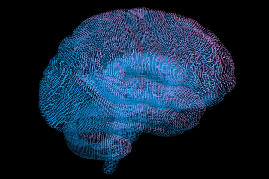
A number of imaging tools are available to study connectivity in autism, each providing a slightly different picture of the disorder.

By Amedeo Tumolillo
22 March 2013 | 1 min read
This article is more than five years old.
Neuroscience—and science in general—is constantly evolving, so older articles may contain information or theories that have been reevaluated since their original publication date.

A number of tools are available to study connectivity in autism, each with strengths and limitations.

Structural magnetic resonance imaging (MRI) takes static pictures of the brain that researchers can use to measure the volume and thickness of the structures within. It can also resolve the corpus callosum, the long and thick bundle of fibers that bridges the left and right hemispheres of the brain (shown here with its seven subsections in different colors). The corpus callosum is smaller in individuals with autism than in controls. Structural MRI can also measure the total volume of white matter, the nerve fibers that connect different regions, but it cannot detect the vast majority of white matter tracts in the brain.