Research image of a mouse brain
Quiet time: Most brain regions are responsive to the new pPDH marker, but the hippocampus and some regions outside the blood-brain barrier are not.
Courtesy of Yu Wang, Dong Yang and Li Ye

What goes up must come down: New marker flags decreased neural activity

Phosphorylation of the metabolic enzyme pyruvate dehydrogenase inversely correlates with neural activity, offering scientists a tool to study inhibition for the first time.

Phosphorylation of the metabolic enzyme pyruvate dehydrogenase inversely correlates with neural activity, according to a new study. This marker of decreased neural activity — the first ever described — doubles the data available to researchers who have long used c-FOS and other “immediate early genes” to pinpoint increased neural activity.

The phosphorylated pyruvate dehydrogenase (pPDH) marker fills a gap in basic neuroscience research, says lead investigator Li Ye, professor of neuroscience at the Scripps Research Institute. “The existing markers — the [immediate early genes] and the c-FOS — only tell you when the neurons are activated; and if the neurons stop being activated, or they are inhibited, that doesn’t report.”

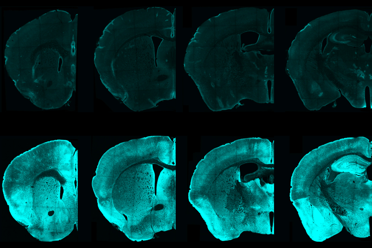
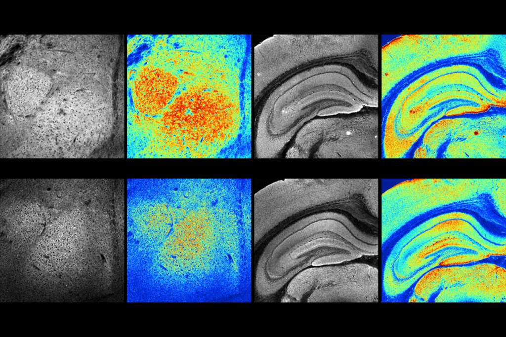
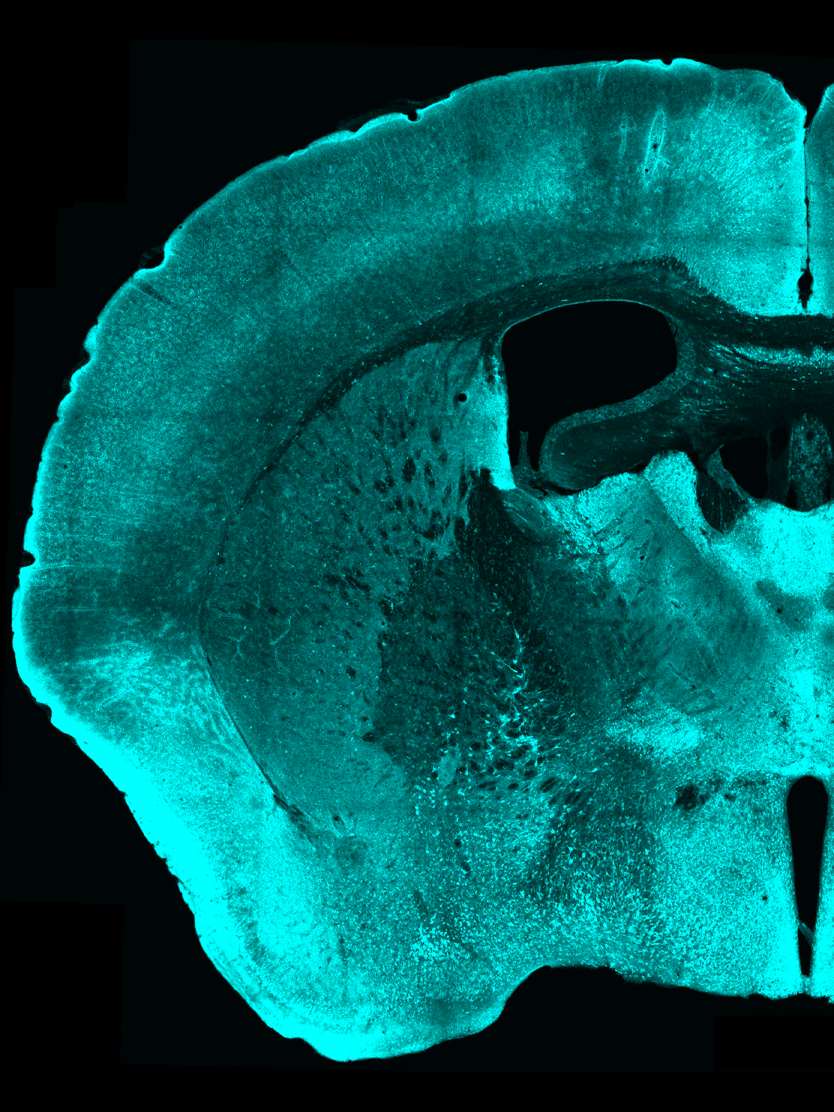
Now, washing fixed tissue slices with antibodies that target pPDH can light up those inhibited cells. By way of example, the team tested the method in mice temporarily lacking access to water and identified 10 brain regions previously unknown to be regulated by thirst.

Scanning for decreased neural activity can illuminate regions previously unidentified by c-FOS or other activity markers, says Vineet Augustine, assistant professor of neurobiology at University of California, San Diego, who advised the team on the thirst experiment. “This is a more holistic approach to looking at behavior readouts.”

T

he discovery, which took more than five years of work and combined optogenetics with proteomics screening, fulfills only half of Ye’s original goal: to identify a single marker that can track both the increase and decrease of neural activity.

One key tool behind the breakthrough, Ye says, was an ultrabright LED light with enough power to stimulate action potentials in millions of primary cortical neurons transduced with channelrhodopsin. He and his team used the LED to make plates of cells fire in synchrony at either 0.5 hertz or 10 hertz. Afterward, they lysed the neurons and hunted for proteins that differed between the slow- and fast-firing groups.

Only one enzyme emerged: pPDH, which is involved in mitochondrial metabolism.

PDH is one of three subunits in the pyruvate dehydrogenase complex that kickstarts the Krebs cycle to produce energy for the cell in the form of ATP. When the PDH subunit is phosphorylated, though, ATP production stops. Low neural firing — which requires less ATP — might correlate with high levels of pPDH, the authors hypothesized.

Coincidently, a commercial antibody matching pPDH became available soon after the team flagged it as a potential marker, Ye says. Antibody in hand, the team verified that pPDH levels increased after they suppressed spontaneous neural activity in cultured neurons and in mice following anesthesia. The marker lit up nearly all regions of the anesthetized mouse brain.

It was at this point that study investigator Dong Yang, also at the Scripps Research Institute, says he realized that the tool would “attract the interests of many scientists who focused on different brain regions.”

Proving that pPDH correlates with behavior was more difficult, Ye says. “The cells don’t get inhibited when you do the opposite things to activation.”

Research image of mouse brains
Lighting up: The marker pPDH picks up suppressed brain activity in mice two hours after general anesthesia (bottom row), but not in control brains (top row).
Courtesy of Yu Wang, Dong Yang and Li Ye

Moving mice from light to dark, for example, did not result in immediate pPDH increases, Ye says. Instead, mice placed in darkness for just two hours following a bright light exposure to trigger neural activity showed more pPDH in their brains than mice that acclimated to the darkness over many more hours. The pPDH caught only the reduction of light-induced neural activity, the team wrote in their paper, published in Neuron on 23 January.

pPDH also correlates with decreased activity in the brain region associated with fasting soon after the mice regain access to food, the researchers found. The thirst experiment they described in the paper suggests the new marker acts more closely in time with neural activity than do immediate early genes such as c-FOS.

F

or all its promise, pPDH has limitations: It does not label regions outside the blood-brain barrier or neurons in the hippocampus, the researchers reported. It is also limited to postmortem tissue, has been tested only in mice and calls for behavioral tests occurring on long time scales — an hour or more.

Some researchers who have already used the marker have also asked Ye about the messiness they see when they stain pPDH. Unlike c-FOS, which stains in the nucleus, pPDH is in the cytoplasm of the dendrites, axons and cell bodies. In regions where pPDH staining ends up looking like “spiderwebs,” Ye says, “you can’t just count cells; you have to find a different way to quantify them.”

Amar Sahay, professor of psychiatry at Harvard Medical School, who was not involved in the study, says he would have liked the researchers to directly manipulate inhibitory neurons and label their downstream connections with pPDH. “That is harder to do when you have to sacrifice the mice,” he admits. “But it still allows us to visualize as a direct consequence of recruiting a specific inhibitory neuron how the cells in the target field respond.”

But already, with no additional hardware needed, this tool can quickly identify brain regions controlled by inhibition, something that is “almost impossible to do by calcium imaging,” says Christina Kim, assistant professor of neurology at University of California, Davis, who was not involved in the study. It can also easily combine with other stains or proteomic profiling techniques to find correlations across brain regions, she adds.

Last March, just as Francesco Roselli, professor of neurology at Ulm University, was searching for a way to measure inhibition in spinal cord circuits, a preprint of Ye’s study appeared on bioRxiv. Roselli, who was not involved in the work, seized on it. The process was straightforward and easy to implement, he says, and soon the commercial antibodies for pPDH lit up the inhibitory synapses he studies for his work on neurodegeneration.

More inhibitory markers will come soon, Roselli predicts. “We are parking in an empty parking lot — there are plenty of spots left open.”

Where do you want to use the pPDH marker to study suppressed neural activity? Leave a comment below.