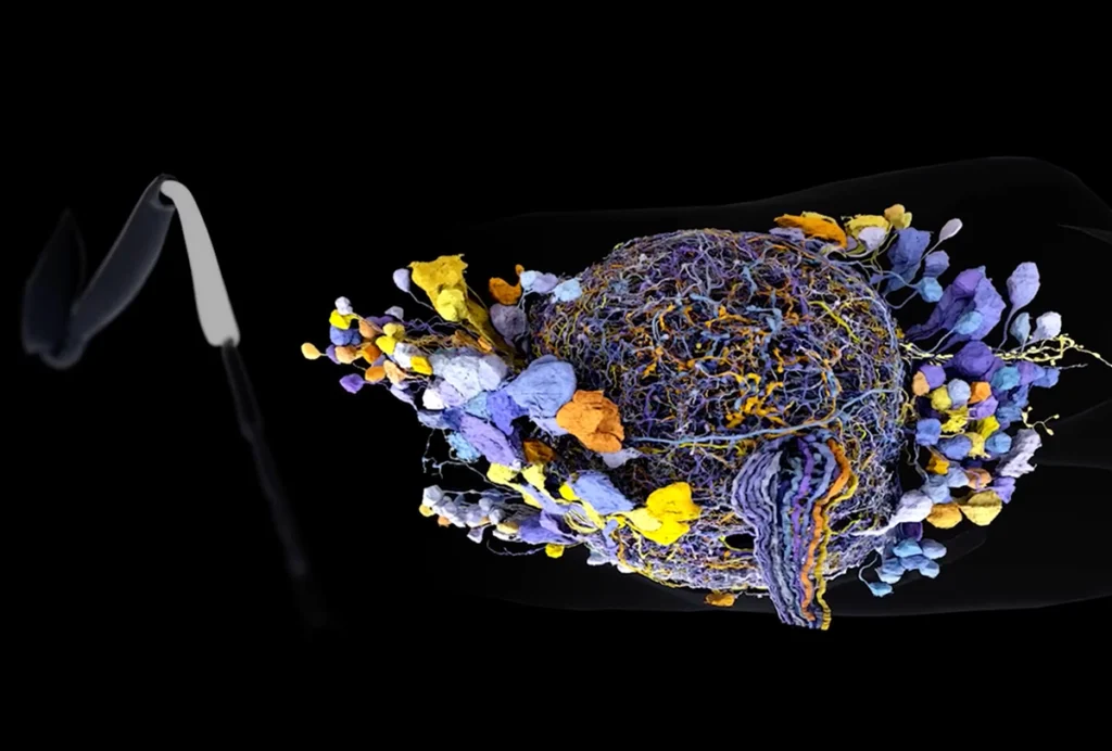
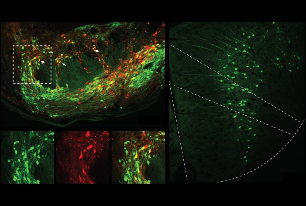
Research video of a zebrafish larva (zoomed in on the gut) being given glucose.
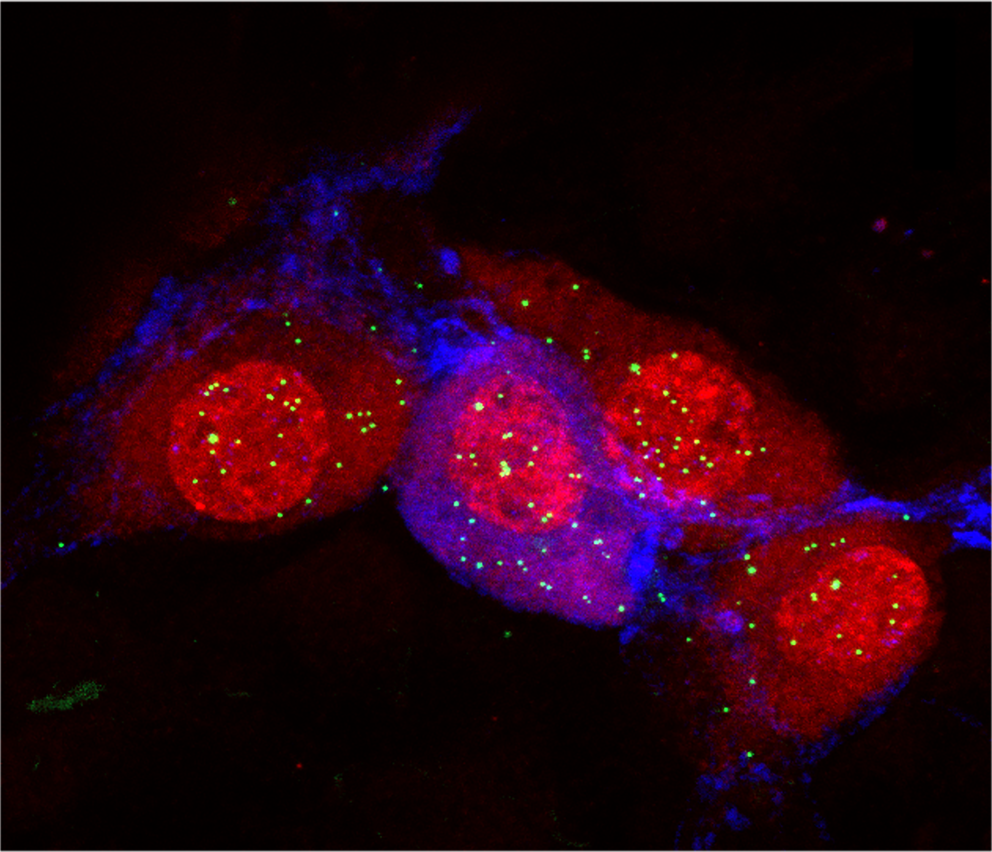
Sluggish flow: Zebrafish larvae lacking the autism-linked gene SHANK3 have fewer enteroendocrine cells in the gut wall and slower digestion than wildtype fish, echoing the severe constipation often seen in children who carry a mutation in SHANK3.
Courtesy of Elizabeth Davidson / Dallman Lab

On the periphery: Thinking ‘outside the brain’ offers new ideas about autism

Neuronal alterations outside the brain may help to explain a host of the condition’s characteristic traits, including sensory changes, gut problems and motor differences.

M

any papers about autism-linked genes note that the genes are expressed throughout both the central and the peripheral nervous systems. The proportion of such prolific genes may be as high as two-thirds, according to one 2020 analysis. Yet few studies delve into what those genes are actually doing outside the brain.

That’s starting to change. Although autism is typically thought of as a brain condition, a critical mass of researchers has started to investigate how the condition alters neurons elsewhere in the body. Their work — part of a broader trend in neuroscience to look beyond the brain — hints that the role of the peripheral nervous system in autism is, well, anything but peripheral: Neuronal alterations outside the brain may help to explain a host of the condition’s characteristic traits.

Much of the research so far focuses on touch and the workings of the gut, but there is increasing interest in other sensory and motor neurons, as well as the autonomic nervous system, which orchestrates basic body functions such as heartbeat, blood pressure, breathing and digestion.

It’s difficult to pinpoint whether some autism traits arise in the peripheral nervous system or the central nervous system; in many cases, complex feedback loops link the two. “Your nervous system doesn’t know that we’ve divided it that way,” says Carissa Cascio, associate professor of psychiatry and behavioral sciences at Vanderbilt University in Nashville, Tennessee.

But at least some peripheral changes may offer novel treatment targets. And drugs that act in the peripheral nervous system could also prove more effective and have fewer side effects than brain-based therapies, says Julia Dallman, associate professor of biology at the University of Miami in Coral Gables, Florida.

“I think there’s actually a lot of opportunities for peripherally targeted treatments,” says geneticist Lauren Orefice, assistant professor of genetics at Harvard Medical School and Massachusetts General Hospital. “It’s interesting because it is the opposite of what we’ve tried to do in neuroscience for a really long time” — that is, getting drugs into the brain.

This new focus on the periphery is already prompting fresh thoughts about old data, says Elisa Hill-Yardin, a neuroscientist at RMIT University in Melbourne, Australia. When she set out to investigate the role of autism-linked genes in the gut, for example, the NLGN-3 mouse was one of few autism mouse models available. When she discovered gut problems in the mice, she reached out to the doctors who had cared for the first children identified with NLGN-3 mutations.

“And lo and behold, those two boys, who are now adults in Sweden, both had really quite serious gastrointestinal dysfunction,” Hill-Yardin says. The details had “been beautifully recorded” by the doctors but went unmentioned in the report because they seemed irrelevant in characterizing an autism-related gene.

Here we take you on a quick tour of some of the different lines of evidence linking autism to the peripheral nervous system.
Touch receptors:

M

any people with autism have unusual responses to touch. Some are hypersensitive to the slightest tap; some are soothed by the constant, all-over pressure of a weighted blanket.

Alterations to a diverse array of sensory receptors may drive such atypical responses. For example, mice lacking the autism-linked genes MECP2GABRB3 or SHANK3 only in certain touch neurons are hypersensitive to puffs of air on their back, researchers led by David Ginty, professor of neurobiology at Harvard University, reported in 2016.

“When we deleted these genes in the peripheral sensory neurons, that essentially perfectly recapitulated the tactile hypersensitivity we observed in mice that had the genes deleted throughout the entire body, including the brain,” says Orefice, who conducted the work as a postdoctoral researcher in Ginty’s lab and is continuing the investigations in her own lab. By contrast, disrupting one of the genes, MECP2, only in a part of the brain responsible for processing sensory information did not alter the animals’ responses.

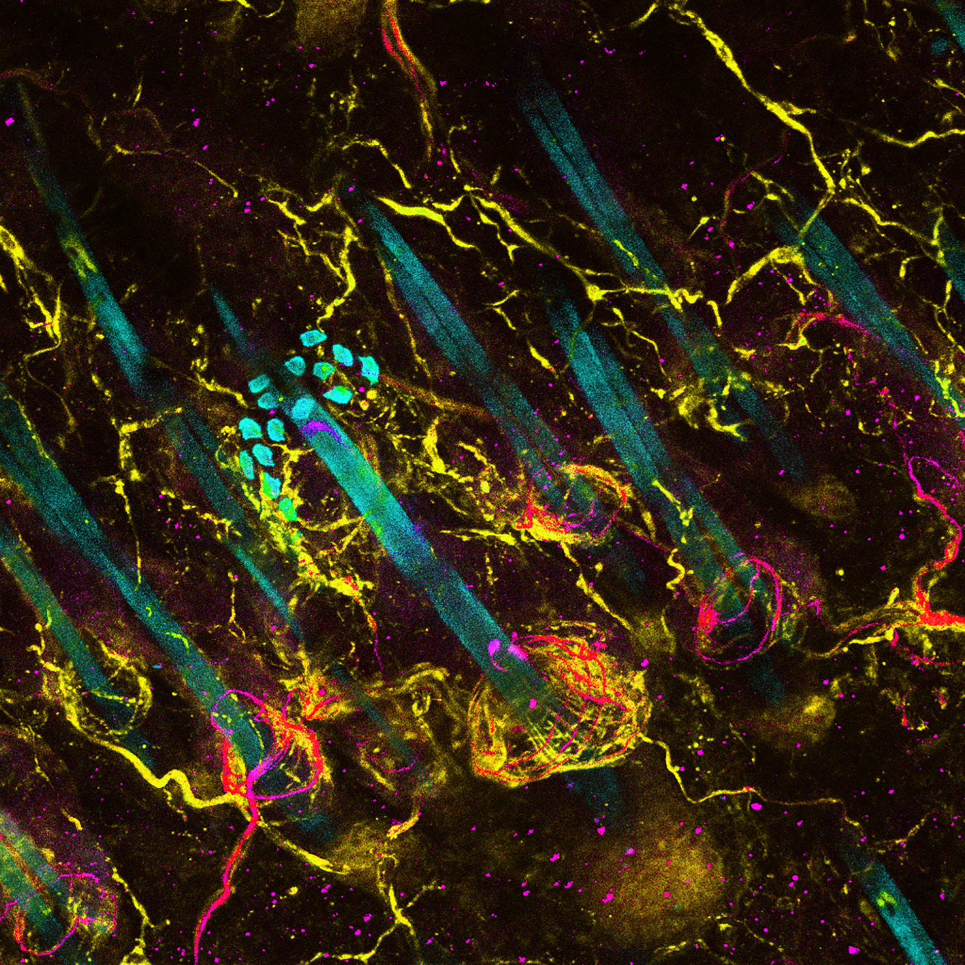
Research image of sensory neuron endings wrapping around hair follicles in a mouse’s skin.

On the surface: Mice missing autism-linked genes only in their peripheral sensory neurons, shown wrapped around hair follicles, have anxiety and other autism-like traits.

Courtesy of Lauren Orefice


Mice missing the genes in touch receptors from birth also display anxiety and autism-like social differences, the team found. But if the genes are turned off in the touch receptors later in life, the animals have tactile hypersensitivity but not anxiety, and only mild social impairments.

“So, a deletion in the peripheral neuron is actually leading to changes in brain-driven behaviors,” Orefice says. The finding suggests that when peripheral nerves don’t function typically, it may change how social areas of the brain develop — and in so doing, drive some of autism’s core traits.

Drugs that dampen the activity of touch receptors can ease touch hypersensitivity, anxiety and some social deficits in six different mouse models of autism, Orefice and her colleagues showed in another study, raising the possibility that drugs that act in the peripheral nervous system might relieve sensory overload and perhaps other difficulties for people with autism.

Further reading:

C-tactile fibers:

O

ver the past several years, researchers have homed in on a particular class of touch receptors called C-tactile fibers that may have particular relevance to autism.

These peripheral neurons are thought to be especially important in so-called social or affective touch: gentle touch with an emotional component that cements social bonds, such as a cuddle from a parent, a hug from a friend or a caress from a romantic partner.

People with autism sometimes find affective touch unpleasant, leading to speculation that the condition can involve atypical C-tactile fibers. Skin samples from four autistic children, for example, contained about half as many C-tactile fibers as is typical, one study found. There is relatively little direct evidence so far. But a recent mouse study bolsters the argument.

Mice genetically engineered to have less-active C-LTMRs — the mouse equivalent of C-tactile fibers — are less sociable than wildtype mice, according to the study, in which researchers used an automated system to track the animals’ behaviors. And temporarily enhancing the function of C-LTMRs in another group of mice increased their sociability, the team found.

“By targeting the peripheral nervous system, you can impact social behavior,” says study leader Amaury François, a neuroscientist at the Institute of Functional Genomics at the French National Center for Scientific Research in Montpellier. “Stimulation of the C-LTMR in mice is pleasant and gives rise to prosocial behavior. And on the other side, reducing the activity of the C-LTMR reduces the seeking of social interaction.”

François and his colleagues are now investigating whether lowering C-LTMR activity early in life affects social behavior in adult mice. They also plan to examine C-LTMR and other touch-receptor function in mouse models of autism.

Further reading:

Pain receptors:

C

-tactile fibers make up a small proportion of C-fibers. Most of the skin’s C-fibers are pain receptors, also called nociceptors.

Many people with autism have an unusual response to pain; they can be either oversensitive or undersensitive to painful stimuli. And autism-linked genes can affect the function of pain receptors in the skin, according to several studies in mice.

Mice missing one or both copies of SHANK3 — implicated in Phelan-McDermid syndrome, an autism-related condition that involves reduced sensitivity to pain — are less sensitive to pain than wildtype mice are, one 2016 study showed. Mice lacking one or both copies only in sensory receptors in the skin have a similar phenotype, suggesting that disruption of SHANK3 in the peripheral nervous system may be responsible for altered pain sensitivity in people with Phelan-McDermid syndrome.

Mice lacking another autism-linked gene, CNTNAP2, are hypersensitive to pain, a separate study showed two years later. Evidence that immune molecules targeting CASPR2, the protein encoded by CNTNAP2, contribute to some cases of chronic pain inspired the work. Injecting these immune molecules into mice rendered the animals hypersensitive to pain, says study leader John Dawes, associate professor of neurophysiology at the University of Oxford in the United Kingdom.

The molecules did not enter the brain or spinal cord of the mice, suggesting they act by disrupting the function of peripheral neurons instead. Confirming this suspicion, sensory neurons in the skin express CNTNAP2, and these neurons — especially pain receptors — are hyperexcitable in mice lacking the gene, the researchers found.

For Dawes, who is primarily a pain researcher, the findings were reassuringly expected. But “if you come from more of an autism angle, then I think the results can be quite surprising,” he says. “From this model of autism, we showed that there was actually dysfunction in the peripheral nervous system.”

Dawes and his team aim to investigate whether overexpressing CNTNAP2 can dampen pain sensitivity in mice. His team has developed mice in which CNTNAP2 can be inactivated or overexpressed only in specific neurons, which “maybe people in the autism field might want to use,” he suggests.

Further reading:

Odor and chemoreceptors:

S

mell tends to be an underappreciated sense among humans. But people do use their sense of smell to gather clues about others’ emotions. And there’s evidence that autistic people respond differently to these social chemosignals, perhaps because of altered function of olfactory receptors or other aspects of the peripheral nervous system.
In a 2017 study, for instance, researchers exposed 20 autistic men and 20 neurotypical men to two different odors while the men viewed images of faces conveying different emotions. One odor was “fear sweat” collected from skydivers, and the other came from people walking calmly. The participants weren’t consciously aware of what they were smelling, but the fear sweat caused an increase in skin conductance only in the neurotypical participants.

Skin conductance is a measure of electrical activity that indicates activation of the sympathetic nervous system, a branch of the autonomic nervous system involved in the fight-or-flight response. The lack of response to the fear odor among the autistic participants suggests that their olfactory receptor function may be altered, the researchers say.

Animal findings lend further support to the idea that autism-linked genes affect the function of receptors used for chemosensation. In fruit flies lacking the autism-linked gene NF1, male flies attempt to court other males — altered social behavior that cannot be reversed by restoring NF1 in the central nervous system.

“We couldn’t fix things, no matter where we were putting it in the brain,” says study leader Matthew Kayser, associate professor of psychiatry at the University of Pennsylvania Medical School in Philadelphia. “So we started saying, ‘Gosh, is it possible that if we put this back in peripheral neurons that it would fix the problem?’”

Kayser’s team was able to stop the fruit flies from courting other males by restoring NF1 in their gustatory sensory neurons. These neurons, found on the surface of the fly’s body, detect chemical cues that would normally signal that another fly is male and inhibit courtship. Such neurons do not exist in people. “It was a real change in how we thought that this problem was arising,” Kayser says.

Further reading:

Peripheral neurons in the gut:

P

eripheral neurons in the gut, which make up a branch of the autonomic nervous system known as the enteric nervous system, regulate contraction, secretion, absorption and other processes in the digestive system — and can do so independently of the brain. In mouse studies in which gut tissue is cultured in laboratory dishes, “you can just record using a video — these beautiful, spontaneous contractions by the gastrointestinal tract that are regulated by the enteric nervous system,” Hill-Yardin says.

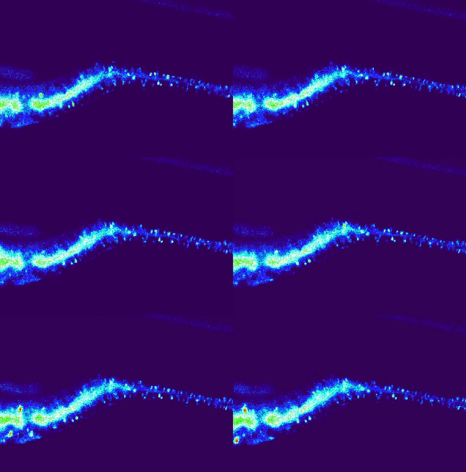
Research image of neurons in the small intestine of a wildtype mouse.

Gut check: The small intestine in wildtype mice has fewer neurons (shown here) than that in mice with an autism-linked NLGN3 mutation — animals that also have digestive issues similar to those autistic people experience.

Courtesy of Herath, Hill-Yardin et al. / RMIT University and The University of Melbourne, Australia


Using just such an experimental setup, Hill-Yardin and her colleagues demonstrated that gut tissue from mice carrying an autism-linked mutation in the gene NLGN3 is more sensitive to drugs that mimic the signaling molecule gamma-aminobutyric acid (GABA) — a result that other researchers had previously shown in brain slices taken from the mice.

Mice with the NLGN3 mutation also have an increased number of neurons in their small intestines, and food moves through the small intestine faster than it does in wildtype mice, Hill-Yardin and her team reported in 2019.

NLGN3 is just one of more than half a dozen autism-related genes that have been linked to altered gut function in recent years — findings that suggest chronic constipation, diarrhea, reflux and other gut problems common among autistic people are more than curious comorbidities or medication side effects.

Similarly, mice lacking one copy of the autism gene FOXP1 also have altered gut function. “We found a really strong alteration which may explain some of the features that the patients [who carry FOXP1 mutations] have,” says study leader Gudrun Rappold, a geneticist at the University of Heidelberg in Germany. The mice have an impaired ability to push food from the esophagus into the stomach and lowered contractility of the colon, so that food takes longer to pass through the gut than it does in wildtype mice. What’s more, genes regulated by FOXP1 in the brain also are dysregulated in the animals’ gut tissue.

Rappold and University of Heidelberg geneticist Beate Niesler reviewed studies of 62 genes strongly linked to autism and found that more than 90 percent are expressed in the gut as well as the brain. But in many cases, it’s not yet clear whether these genes are actually expressed in enteric neurons or whether alterations in gut function can be traced to enteric neurons specifically.

The enteric nervous system is the “dirty end” of the nervous system, Hill-Yardin says: Relatively little is known about it compared with the brain, and tools to explore it and background knowledge are often lacking. But this is beginning to change. Researchers have put together a set of tools to manipulate gene expression in particular populations of gut neurons, for example.

In the past, much research on gut problems in autism focused on suspected differences in the microbiome between autistic people and controls. But Hill-Yardin and her collaborators showed that NLGN3 mice have altered microbiota compared with wildtype mice housed in the same cage, suggesting that altered autism-linked genes could even drive gut microbiome changes in autistic people. Other researchers theorize that altered function of the autonomic nervous system may change the biology of the gut, including reshaping the microbiome.

Further reading:

Enteroendocrine cells in the gut:

A

s is true for FOXP1 mice, food moves more slowly through the gut of zebrafish larvae lacking one copy of SHANK3 than it does in wildtype fish, echoing the severe constipation often seen in children who carry a mutation in SHANK3. Researchers have traced this abnormality in the fish to a loss of enteroendocrine cells in the gut wall.

Enteroendocrine cells, as their name implies, release a variety of hormones and are conventionally thought of as part of the endocrine system. But the boundaries of the nervous system can be fuzzy: These cells also release neurotransmitters, including serotonin and glutamate, and have processes that protrude into the lumen of the gut to gather information on the gut’s contents, which they then relay to the brain.

“The enteroendocrine cells clearly have neuronal aspects to them,” says study leader Dallman.

Some enteroendocrine cells are chemosensitive and signal for the gut to slow its contractions when they detect sugar or fat, to aid absorption of these nutrients. Others are mechanosensitive and detect when a bolus of feces needs to be moved along and out of the body. “They are basically the sensory cell of the gut,” Dallman says.

These mechanosensitive enteroendocrine cells are the ones that are most affected in the SHANK3 fish, Dallman and her team found. “It means that there’s less of a way of conveying what’s going on in the lumen of the gut to the rest of the organism,” she says. That lowered capacity again maps to the constipation that plagues many people who carry a mutation in SHANK3.

Dallman and her team plan to conduct experiments on gut tissue in laboratory dishes to confirm that the altered gut function arises from the gut rather than the brain in the SHANK3 fish, and they also have unpublished data implicating enteroendocrine cells in gut problems in fish lacking another autism-linked geneSYNGAP1.

They are also investigating whether using optogenetics to turn on enteroendocrine cell function can relieve gut problems in the SHANK3 fish. If so, the cells could be a target for treatment to reduce constipation in people.

Further reading:

Interoception and baroreceptors:

T

he most familiar sensory receptors are ones that gather information about sights, smells, tastes and tactile sensations from outside the body. But there are also sensory receptors that monitor processes inside the body, contributing to a sense known as interoception. Such receptors include the baroreceptors that enable people to sense their own heartbeat, for example.

Data from Cascio’s lab indicate that some people with autism struggle to discern whether or not their heartbeat is aligned with a pulse of light or sound. This finding suggests that some autistic people have trouble integrating internal and external signals, which could contribute to anxiety, Cascio says.

It’s not yet clear whether these differences arise in the sensory receptors that gather internal information or in the brain regions that process this input. But in brain imaging studies, Cascio has found no differences between autistic and neurotypical people in activity in the brain areas that process interoceptive information. Given those findings, “it’s logical to think that there might be more of a peripheral difference,” Cascio says.

Autistic people tend to be more aware of a broad array of internal sensations than neurotypical people are, according to questionnaires. This heightened awareness could contribute to the feelings of sensory overwhelm that many autistic people report, says Lisa Quadt, neuroscience research fellow at the University of Sussex in the U.K.

Quadt and her colleagues have found evidence that non-pharmacological treatments to improve awareness and interpretation of interoceptive signals could help ease some of the difficulties autistic people encounter in their daily lives. In one of the largest randomized trials for anxiety in autistic adults yet conducted, the researchers tested such an intervention, called Aligning Dimensions of Interoceptive Experience (ADIE), designed in consultation with autistic people.

After six weeks, 60 autistic adults who received ADIE — including providing them with feedback about the accuracy of their heartbeat counts — saw a decrease in their background levels of anxiety, compared with 60 autistic adults who didn’t get the intervention. What’s more, participants’ scores on a questionnaire that broadly measures how aware people are of various body signals went down.

“There might one item in these 45 questions that asks about your heartbeat, but [the questionnaire is] about what is happening in your entire body,” Quadt says. “So I found this so great, that this one channel that we improve — the heartbeat that got more accurate — had this effect on the whole body.”

“There was a participant who put this into beautiful words,” Quadt adds: “‘The more precise my inner signals got, the less overwhelming the outer world got.’”

Further reading:

Peripheral motor neurons:

A

lterations in autism-related genes may also affect the function of motor neurons, which communicate with muscles to orchestrate body movements. By studying mice lacking SHANK3 and tissue from people with Phelan-McDermid syndrome, researchers established that the lack of SHANK3 disrupts development of motor neurons, neuromuscular junctions and skeletal muscles.

“The whole motor system is altered,” says study leader Tobias Böckers, a neurobiologist at the University of Ulm in Germany. These insights could help explain why people with Phelan-McDermid syndrome have low muscle tone, and they hint at the potential involvement of the peripheral nervous system in motor problems in autism more broadly.

In a follow-up study, the researchers showed that SHANK3 is also expressed in oligodendrocytes and Schwann cells, which are responsible for the formation of myelin, the insulating sheath around neurons that helps signals transmit quickly. Mice lacking SHANK3 have reduced levels of myelin proteins in the central nervous system but increased levels in the peripheral nervous system, suggesting that the speed of information transmission and processing may be altered in complex ways.

In turn, the findings suggest that the characteristics of Phelan-McDermid syndrome may not be solely due to a neurodevelopmental problem in the brain but to an interplay between altered input from the periphery and disrupted brain development. “It makes the story a lot more complicated” than the conventional brain-centric view of neurodevelopmental conditions, Böckers says.

Further reading: