After the publication in 1992 of a seminal paper detailing insights from functional MRI, scientists focused for two decades on comparing scans from large groups of people. But in 2012, a researcher named Russell Poldrack introduced a new approach to the field.
Poldrack wanted to know more about the individual brain rather than group averages generated by large-scale scanning projects. He was aware of volume fMRI scanning in the vision neuroscience field, and he knew animal models had shown that long-term exposure to high magnetic fields didn’t carry a significant biological effect. But he couldn’t figure out how to conduct repeated fMRI scanning on a volunteer: The time commitment and frequent trips to the lab seemed like a big ask.
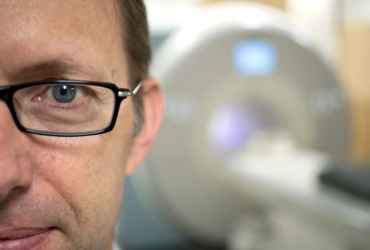
So, Poldrack says, “I figured that before I try to do it on someone else, I should do it with myself. Because I knew I would be the best subject I was going to be able to find.”
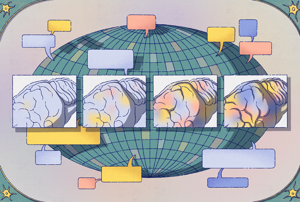
Poldrack’s serial scanning of his own brain kicked off a mini-movement. Today, there is an increasing number of researchers who have published papers based on precision scanning—putting one person in the fMRI machine over and over again—which are revealing important differences among individual brains. Almost all of them owe a debt to Poldrack’s first study on himself. His way of solving the commitment problem associated with precision scanning proved to be a contagious workaround, and the concept jumped from one lab to another over the years, surprising even its creator.
“I had no idea that it would take off in the way that it has taken off, for sure,” Poldrack says.
The vignettes below trace how Poldrack’s innovation moved from lab to lab, from one scientist to another.
On the afternoon of 24 September 2012, Poldrack went to the basement of his research building at the University of Texas at Austin and slid into the MRI scanner. He was professor of psychology and neurobiology and director of the Imaging Research Center at the university, and he had been frustrated with the state of cognitive neuroscience for some time.
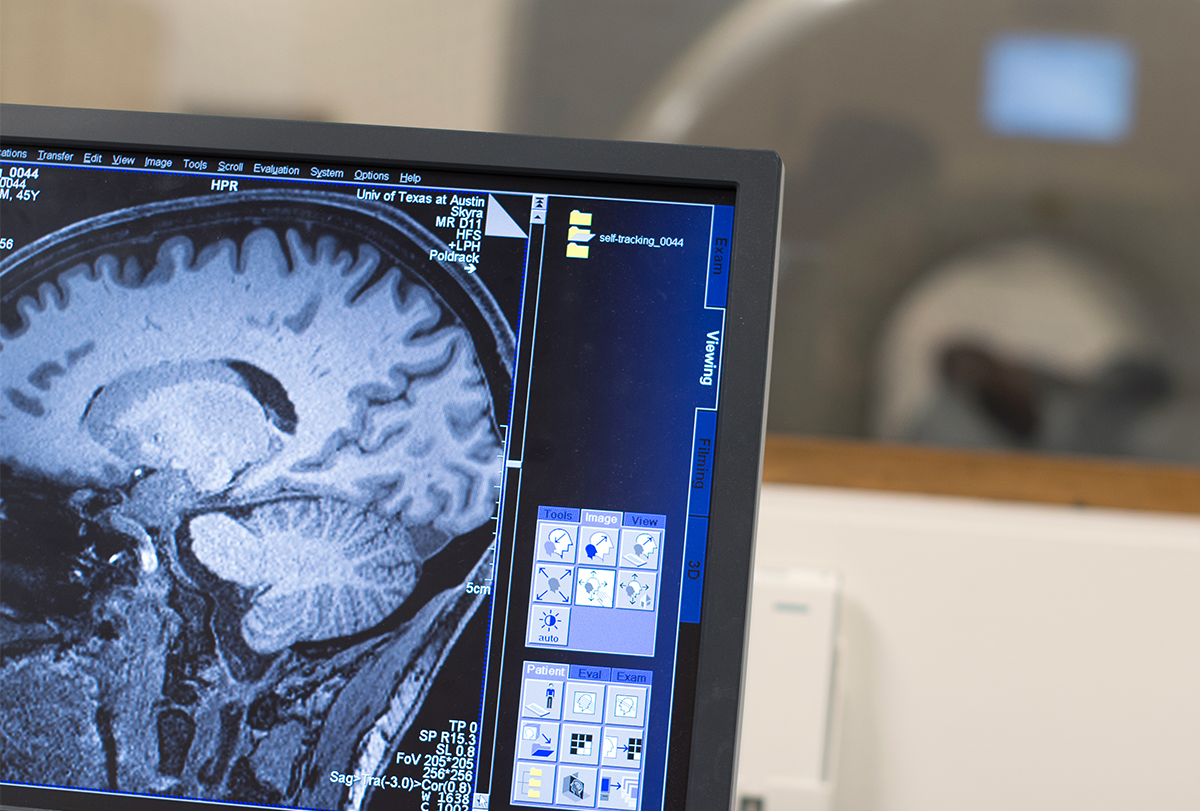
Single source: Over 18 months, Poldrack scanned his own brain more than 100 times.
Courtesy of Russell Poldrack
For years his field had been enthralled with fMRI as a way to better understand neural networks, but Poldrack felt scans captured only “a little bit of data from each person.” His psychology background had taught him that the severity of mental health conditions can vary from day to day, and he felt science needed to know how even a healthy brain fluctuated over time.
Yet no one had ever scanned a brain daily before, as far as Poldrack knew. That meant that any long-term side effects were unknown, and he wondered if his institutional review board would approve a plan to do repeated scans on a volunteer. Plus, he doubted he could convince anyone to commit the time and effort required to participate.
An artist friend suggested that Poldrack simply scan himself, but Poldrack resisted—it felt “pretty narcissistic,” he says. But then he saw Michael Snyder’s “integrative personal omics profile” study, published in 2012, which assessed Snyder’s genome, transcriptome, proteome, metabolome and auto-antibody profile over 14 months. Poldrack realized “that you can actually get interesting scientific insights out of data from one person.”
So here he was in the belly of the machine. Over the coming 18 months, he would scan himself more than 100 times, while also drawing blood for gene-expression and metabolite levels. The eventual result was “MyConnectome”—a yearlong look at his fluctuating brain, posted online, available for anyone to download and analyze.
The results showed that the brain’s connections are so dynamic they can be linked to gene expression and metabolic function, and the project changed how the neuroscience community thought about brain scanning. Researchers have used data from Poldrack’s project to produce at least 50 academic papers, and Poldrack’s 2015 paper has gathered more than 450 citations. Poldrack, now professor of psychology at Stanford University, has heard from some of these people himself, he says. They approach him at conferences and say, “Hey Russ, come to my poster—it has your brain in it!”
The work’s influence would spread across the neuroscience field, starting with graduate student Timothy Laumann.
In 2012, Timothy Laumann was in an M.D./Ph.D. program for psychiatry and neuroscience at Washington University in St. Louis. He was working on his thesis and mulling over dynamic functional connectivity—it wasn’t clear to him if the networks identified in individual resting-state brain scans would be consistent from one person to the next—and this was on his mind when he sat next to Poldrack at a dinner meeting at Washington University. The two researchers talked about Poldrack’s half-finished self-scan project, and Laumann saw a chance to get answers to some of his own questions.
They agreed to collaborate. The first thing Laumann did was look at Poldrack’s resting-state scans. Previous scans from volunteers had shown group-averaged resting-state networks, which are maps of connections among disparate areas of the brain. It’s what resting-state fMRI aims to measure, and Laumann expected the map of Poldrack’s resting-state networks would resemble the group’s composite map. Yet in one area of Poldrack’s scan, the pattern was completely different from what Laumann had expected to see. This was not, as he first thought, “a hole in Russ’s head” but a portion that didn’t line up with the composite map of that region.
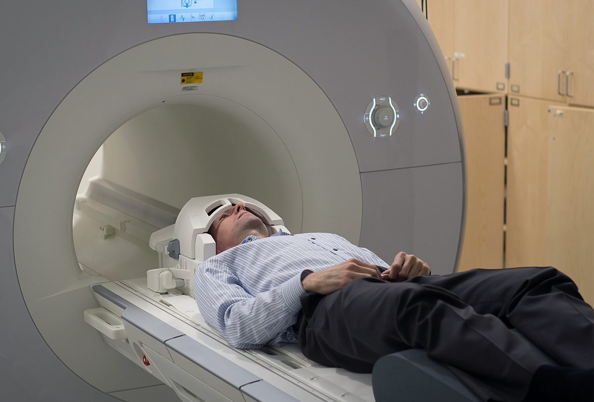
Lie still: Poldrack first climbed into the fMRI machine on 24 September 2012.
Courtesy of Russell Poldrack
Laumann presented the data to his adviser, who told him to run it again. It took several months and multiple analyses, but Laumann finally convinced him. “This is what Russ looks like,” he remembers saying, “and it’s different than everybody else.”
Laumann published his study in 2015, the same year as Poldrack. Laumann’s work showed that, with enough scan data, it was possible to describe the organization of various networks in a single brain—something that had never been done before. The results also showed that distinct features in an individual may differ from group data, and this difference could be vital to understanding psychological and neurodegenerative disorders.
The “Midnight Scan Club” got the idea from working with Laumann.
In 2013, Nico Dosenbach and Steve Nelson were in the early stages of their careers—Dosenbach had just finished his clinical fellowship, and Nelson was nearly done with his postdoctoral research—when they attended a lab meeting in which Dosenbach remembers Laumann presenting prepublication data on his collaboration with Poldrack. Dosenbach recalls being stunned by the work. As he and Nelson walked to Dosenbach’s car, he lamented that he wanted to do something similar but with more participants. He figured it would cost upward of $100,000, and that made it impossible, Dosenbach says, because he “wasn’t the department chair and couldn’t just give myself all the money we needed to do it.”
But Nelson, now associate professor of pediatrics and imaging core director at the University of Minnesota’s Masonic Institute for the Developing Brain, told Dosenbach about a nuance he had seen in Washington University’s scanning costs: They were discounted by 90 percent between midnight and 7 a.m. Dosenbach slapped him on the back. “That makes it doable!” he said. They could get a 10-person dataset for about $10,000.
Because it did not seem reasonable to ask people to repeatedly come to the lab after midnight and lie in a scanner, they decided to “do it ourselves,” Dosenbach says. Similar to Poldrack, becoming the study participant “was from necessity.”
Still, Dosenbach knew they would need to recruit others. “I thought, how do you get people to do something that might not work?” he says. He wanted to create a sense of camaraderie, and he envisioned a motorcycle club. He came up with the Midnight Scan Club and began recruiting members from the broader Washington University neuroscience community. In the end, there was so much interest they had to turn people away.
The study involved 10 participants, each of whom completed 12 scans. The cost ran to just $12,000. That amount for a study is “unheard of,” Dosenbach says, and would typically cover “one [science] meeting with catering.” The Midnight Scan Club’s first paper included 10 detailed, unique functional connectomes and proved that “the group is not a real thing,” says Laumann, who also worked on the paper. “Nobody is walking around with a group brain.”
The club continued. In one standout study, Dosenbach and two other Washington University researchers were scanned before they each had their dominant arm put in a full cast. They were scanned again daily for two weeks while the cast remained in place, and then again after it was removed.
Results showed that after a single day of wearing the cast, brain networks began “decoupling,” says Ashley Nielsen, a cognitive neuroscientist at Washington University and one of the study participants. After the brain had sent signals to the region that controlled the immobilized arm but received back no communication, it began to send pulses to the action mode network instead. When the cast was removed, the brain quickly reverted to the original mapping.
Results like this had been shown in animals but had been difficult to replicate in humans. “The cast paper is among the most important papers in imaging in the last 10 to 15 years,” says Nelson, who was not involved in the study.
The Midnight Scan Club’s work changed the trajectory of Emily Jacobs’ research.
Emily Jacobs, professor of neuroscience at the University of California, Santa Barbara, first read Poldrack’s study and then the work of the Midnight Scan Club. For years, she had taken blood or saliva samples from hundreds of volunteers and then correlated that with one- or two-time fMRI scans of their brains. But hormones and brains change even by the hour, and Jacobs’ group had “never been able to look at these hormones in action and how they’re driving changes in the brain,” she says. Repeated scanning might give them exactly what they were looking for.
Jacobs discussed Poldrack’s work with her lab, and Laura Pritschet, then a 23-year-old Ph.D. student, suggested a day-by-day look at the brain during a young woman’s menstrual cycle. At the time, researchers were split on how fMRI results varied through the menstrual cycles, Pritschet says, because everyone was “doing it differently.”
Pritschet, who is now a postdoctoral fellow in psychiatry at the University of Pennsylvania Perelman School of Medicine, remembers others were hesitant about the idea of repeated self-scanning. “People were like, ‘I don’t know. Should you do that?’” Pritschet says.
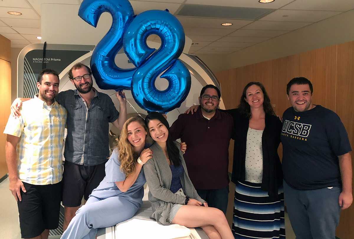
Pritschet forged ahead anyway and offered to be the sole participant in a study. The project—dubbed 28andMe—began not long after the Midnight Scan Club’s initial 2017 paper. Pritschet underwent 30 days of fMRI scans, venipunctures and questionnaires. The result was a never-before-seen glimpse into how the ebb and flow of estradiol, progesterone and other hormones prompt temporary changes in functional networks of the brain.
28 days later: Laura Pritschet (seated on scanner, left), while working in the lab of Emily Jacobs (not pictured), volunteered for 30 days of fMRI scans and other monitoring to detect how fluctuating menstrual hormones affect brain networks.
Courtesy of Laura Pritschet
A year later, the group tested Pritschet while she took a low oral dose of synthetic estradiol as birth control. The results suggested that the brain networks involved in memory and mind-wandering are stronger when estrogen is higher, and weaker when progesterone levels are high.
Then, Pritschet and Jacobs worked with Elizabeth Chrastil to study the effects of pregnancy on the brain, scanning Chrastil before, during and after her pregnancy, and again at 20 weeks, 53 weeks and 122 weeks post-birth. The results showed the real effects pregnancy can have on the brain, and they can last as long as two years—at least in Chrastil.
Most recently, Carina Heller, while visiting the Jacobs’ lab as a postdoctoral fellow with a joint appointment in Bart Larsen’s lab at the University of Minnesota, brought data from her own research project she had completed in Germany. She was one of four women to undergo “dense sampling” across a menstrual cycle. Heller was taking oral contraceptives, and one of the other women in the study had endometriosis. The work is ongoing, but partial results, published last September, showed brain volume changes across the menstrual cycle, and the influence of hormones.
They have begun a larger undertaking called the Longitudinal Menopause Project, with a substudy called the Menopause Scan Club. In addition to Jacobs scanning herself, this time they are also recruiting volunteers. That’s something Harvard University professor Randy Buckner is also doing.
Randy Buckner is professor of psychology and of neuroscience at Harvard University, and he was influenced by Poldrack’s work. “When Russ scanned himself over and over, he was able to make maps across the entire brain with a level of power and precision that the field had not seen before,” Buckner says. “And our lab jumped on it.”
But Buckner didn’t want to scan himself; he wanted outside participants. In his first study, he scanned four people, 24 times each. “It turns out that once people find it easy to do a single session, they don’t have a problem coming back 20 or 30 times,” he says.
Expanding view: Randy Bruckner and his team began a volume scanning project with volunteers performing a memory task. The early data from two participants showed similarities in the individual day to day, but differences between participants
Courtesy of Randy Bruckner
Lauren DiNicola, once a graduate student in the Buckner Lab and now assistant professor of psychology at the University of Virginia, also plans to recruit outside volunteers for her precision-scanning work. Her time working with Buckner showed her it was rare for volunteers to drop out of a study. “Returning to the lab multiple times made them feel like part of this team,” she says, and volunteers wanted to know if they were getting better at remaining motionless during scans, and how the project was progressing. Precision scanning—both self-scanning and in volunteers—in many ways might complement the brain-wide association studies of the past dozen years.
“I am, perhaps naively, optimistic that this is becoming more and more common,” DiNicola says.