Julie Forman-Kay is program head in molecular medicine at the Hospital for Sick Children in Toronto, Canada. She received her B.Sc. in chemistry from the Massachusetts Institute of Technology and her Ph.D. in molecular biophysics andd biochemistry from Yale University. The major focus of her lab is to provide biological insights into how dynamic properties of proteins are related to function and methodological tools to enable better understanding of dynamic and disordered states. Most recently, her lab has probed the biophysics of protein phase separation and how it regulates cellular condensates and biological function.

Julie Forman-Kay
Program head
Hospital for Sick Children
From this contributor

How microscopic ‘condensates’ in cells might contribute to autism
A controversial idea about how cells compartmentalize their contents into droplets — like beads of oil in water — could be key to understanding autism, says Julie Forman-Kay.
How microscopic ‘condensates’ in cells might contribute to autism
By
Angie Voyles Askham
23 February 2021 | 7 min read
Explore more from The Transmitter
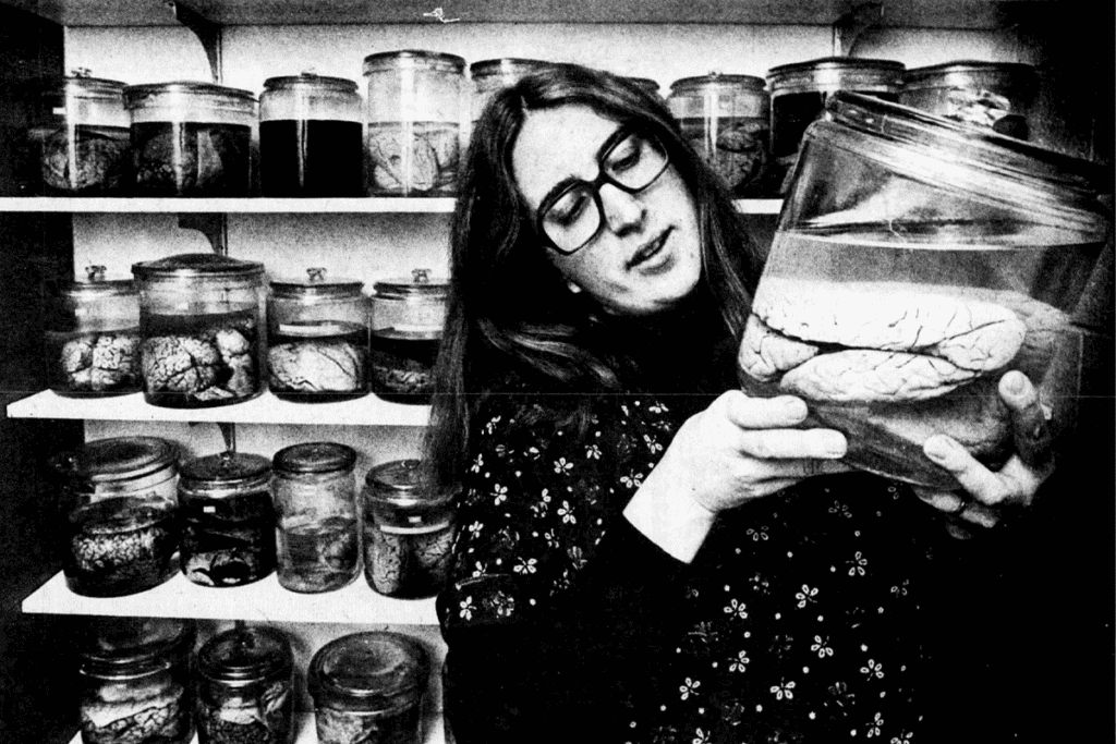
Remembering Barbara Finlay, ‘maverick’ of evolutionary neuroscience
Finlay, who died on 12 September, championed the idea that developmental constraints dictate how evolution sculpts brains, a concept she used to model the timing of hundreds of events and align them across mammalian species.
Remembering Barbara Finlay, ‘maverick’ of evolutionary neuroscience
Finlay, who died on 12 September, championed the idea that developmental constraints dictate how evolution sculpts brains, a concept she used to model the timing of hundreds of events and align them across mammalian species.
By
Calli McMurray
23 October 2026 | 8 min read
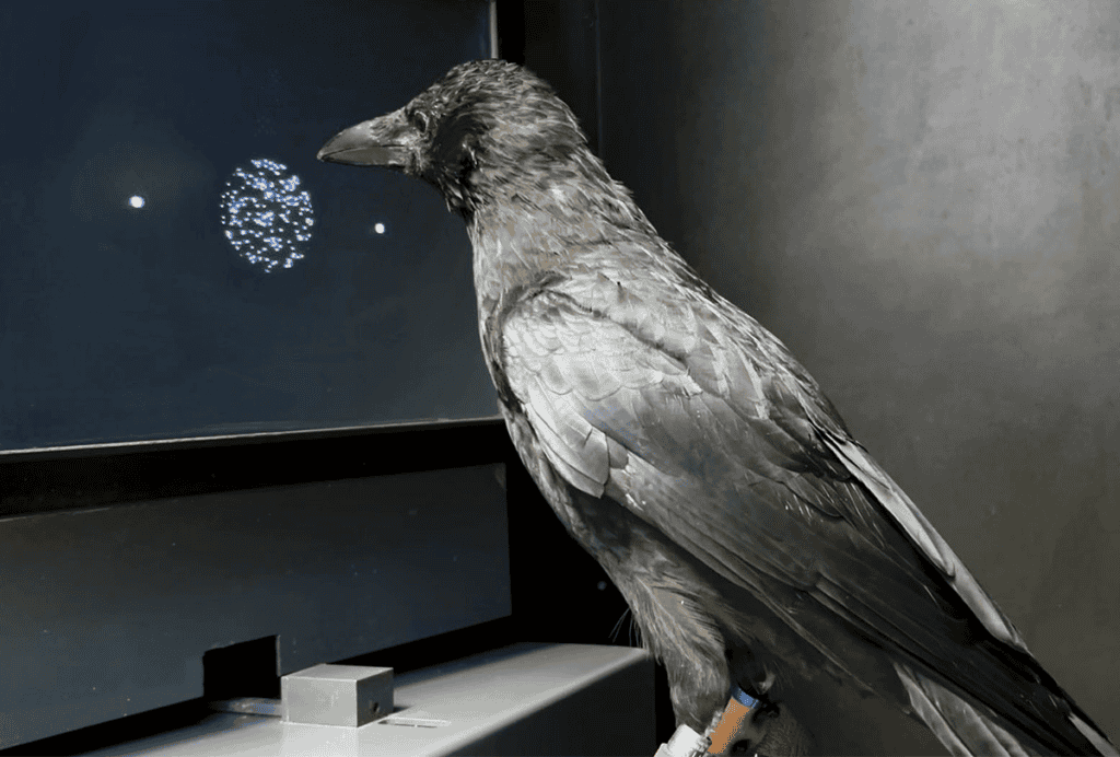
Crows have brains ‘in the fast lane’
The birds accurately complete decision-making tasks twice as fast as primates, although their endbrains look considerably different.
Crows have brains ‘in the fast lane’
The birds accurately complete decision-making tasks twice as fast as primates, although their endbrains look considerably different.
By
Sarah Thau
22 October 2026 | 4 min read

Prevalence of profound autism declines with new definition
The updated, more precise definition used in a CDC study includes adaptive scores.
Prevalence of profound autism declines with new definition
The updated, more precise definition used in a CDC study includes adaptive scores.
By
Brianna Abbott
22 October 2026 | 5 min read