This article is more than five years old.
Neuroscience—and science in general—is constantly evolving, so older articles may contain information or theories that have been reevaluated since their original publication date.
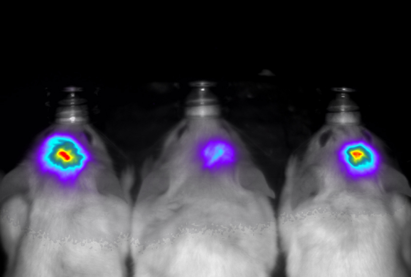
Modified versions of molecules found in fireflies make brain cells glow so brightly they are visible from outside the body.
Modified versions of molecules found in fireflies make brain cells glow so brightly they are visible from outside the body1. The technology may enable scientists to noninvasively track responses of individual neurons inside living animals.
Fireflies’ glow is the result of a reaction between the enzyme luciferase and a compound called luciferin. The resulting yellow light doesn’t penetrate mammalian tissue, however.
The researchers set out to find a version of luciferase that would work with a synthetic molecule called AkaLumine hydrochloride. They subjected the enzyme to several rounds of mutations, and selected the bacterial colonies that fluoresce the brightest in the presence of the molecule. They identified a version of luciferase called Akaluc that produces a bright red glow.
This glow is of a wavelength that can pass through brain tissue, skull and skin. The emission is up to 1,000 times brighter than the firefly reaction, so researchers avoid having to install a clear window or an endoscope to peer inside the animal’s skull.
The researchers injected a virus carrying Akaluc deep into mouse brains, targeting a structure called the striatum that is involved in motor function. When the mice drank water containing AkaLumine hydrochloride, infected neurons in the striatum glowed.
The red glow was visible for an hour while the mice moved freely around their cage.
“We could observe the signals through the intact skull and while the animal is moving naturally,” says lead researcher Atsushi Miyawaki, head of the laboratory for cell function dynamics at the RIKEN Brain Science Institute. Miyawaki’s team measured the fluorescence using a sensitive video camera located outside the cage.
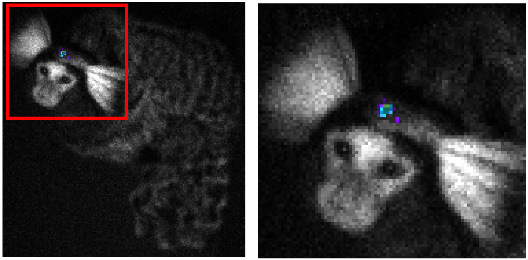
The researchers then injected Akaluc into the brains of mice genetically modified so that the enzyme would be expressed only in active neurons. This time, they targeted the hippocampus, a region involved in memory for places, and put the mice in an unfamiliar container. The bioluminescent molecules revealed an increase in neuron activity in this brain region as the mice explored their new environment.
Analysis of hippocampal tissue in one mouse revealed that only 49 neurons carried the Akaluc enzyme, showing that even a few cells can generate a strong signal.
The researchers also injected a virus carrying Akaluc into the striatum of a female marmoset. Infected neurons in the striatum glowed when the researchers gave the marmoset AkaLumine hydrochloride even a year later. The study appeared 23 February in Science.