Book cover for Bird Brains and Behavior: A Synthesis, showing several bird species.
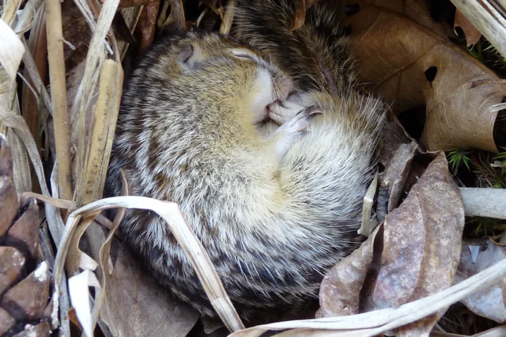
Dozing off: Birds display short bouts of REM sleep, lack a corpus callosum and sleep in only one hemisphere at a time.
Courtesy of MIT Press

‘Bird Brains and Behavior,’ an excerpt

In their new book, published today, Georg Striedter and Andrew Iwaniuk dive deep into the latest research on the neural mechanisms of avian behavior. This excerpt from Chapter 2 explores how birds sleep.

Sleep in birds

Sleep is the most obvious behavior that, in most animals, follows a circadian rhythm. But have you ever seen a bird asleep? Maybe you have, though they usually wake up before you get close enough to see whether they have their eyes closed. Moreover, just because an animal is still and closed its eyes, does that really mean it is sleeping? Maybe it is just resting. Conversely, might some birds sleep with one or both eyes open?

Indeed, it is difficult to tell whether an animal is sleeping just by observing it. To overcome this problem, researchers may prod the animal to see whether it is less responsive at certain times of day. A more definitive method for demonstrating sleep in vertebrates is to record an animal’s brain waves (its electroencephalogram, or EEG), because these waves change significantly as an individual falls asleep and then progresses through several stages of sleep. In birds, the use of EEG recordings is essential because they can sleep with one or both eyes open, presumably so they can stay alert to threats. Ostriches, for example, tend to sleep while sitting on the ground, holding their head up high, and keeping both eyes open. They certainly look alert during this time, but EEG waves reveal that they are actually asleep

Types and patterns of sleep

An EEG measures the activity of many neurons simultaneously. In mammals, it is usually recorded from multiple electrodes placed over the neocortex; in birds, the electrodes are typically placed on top of the hyperpallium (aka the Wulst; see Chapter 1). In addition to performing an EEG, sleep researchers typically record the animal’s eye movements and an electromyogram (EMG), which is a measure of muscle activity, often characterized as muscle “tone.”

These kinds of studies have revealed that, in mammals, the transition from the waking state to sleep is marked by a shift from EEG waves that are low in amplitude (i.e., small) and high in frequency (>20 Hz) to waves that are much larger but lower in frequency (1–4 Hz). Because the latter state is characterized by powerful low-frequency EEG waves (aka slow-wave activity), it is commonly called slow-wave sleep (SWS). The mechanisms that cause SWS are complicated and involve a variety of sleep-promoting processes. However, the large amplitude of these slow waves reflects that, during SWS, numerous neurons fire in rhythm with one another so that their electrical potentials sum when they are recorded through the EEG electrodes.

A fascinating aspect of mammalian sleep is that SWS is occasionally interrupted by periods in which the EEG becomes similar to that of the waking state, even though the animal generally remains motionless, with the notable exception of eye movement (behind closed eyelids). These periods are called rapid eye movement (REM) sleep or, sometimes, “paradoxical sleep”—the paradox being that the EEG indicates wakefulness while other measures reveal the animal to be asleep. It is during this REM sleep that humans experience their most vivid dreams.

No one knows whether birds dream like we do, but EEG recordings from a variety of avian species indicate that birds do have both slow-wave and REM sleep, as well as one or more intermediate states. Several early studies suggested that birds do not sleep very much and usually do so in very short bouts; REM sleep was said to be particularly scarce. However, the experimenters in these early studies left the lights on during the EEG recordings so that they could monitor whether the birds were sleeping or awake. This was unfortunate, because we now know that light severely disrupts avian sleep (which means that city lights can be detrimental to birds; see Chapter 8). If, instead, birds are monitored with infrared light, which is invisible to them, then they sleep much more. Budgerigars, for example, spend 80 percent of their “dark time” asleep, and roughly 30 percent of their total sleep time in REM sleep. Similar results have been obtained from pigeons and songbirds. Altogether, these data show that obtaining a full picture of sleep requires monitoring animals under conditions that are as natural as possible.

Although avian sleep is similar to that of mammals, it does exhibit some significant differences. For one thing, REM sleep episodes tend to be much shorter in birds than in humans. In budgerigars, for example, each period of REM sleep lasts only five to 15 seconds, whereas REM episodes in humans typically last 10 minutes or more. Episodes of SWS also tend to be significantly shorter in birds than in mammals (interrupted as they are by frequent REM). Another species difference is that pigeons dilate their pupils during SWS, whereas mammals constrict them. The opposite happens when the animals are awake: Fully awake mammals tend to have enlarged pupils, whereas aroused birds tend to constrict them. These behavioral differences correlate with a difference in the pupillary constrictor muscles: They are slow-acting, involuntary “smooth muscles” in mammals, but fast, striated muscles in birds. In fact, parrots occasionally communicate by rapidly constricting and then dilating their pupils (a behavior called “eye pinning”).
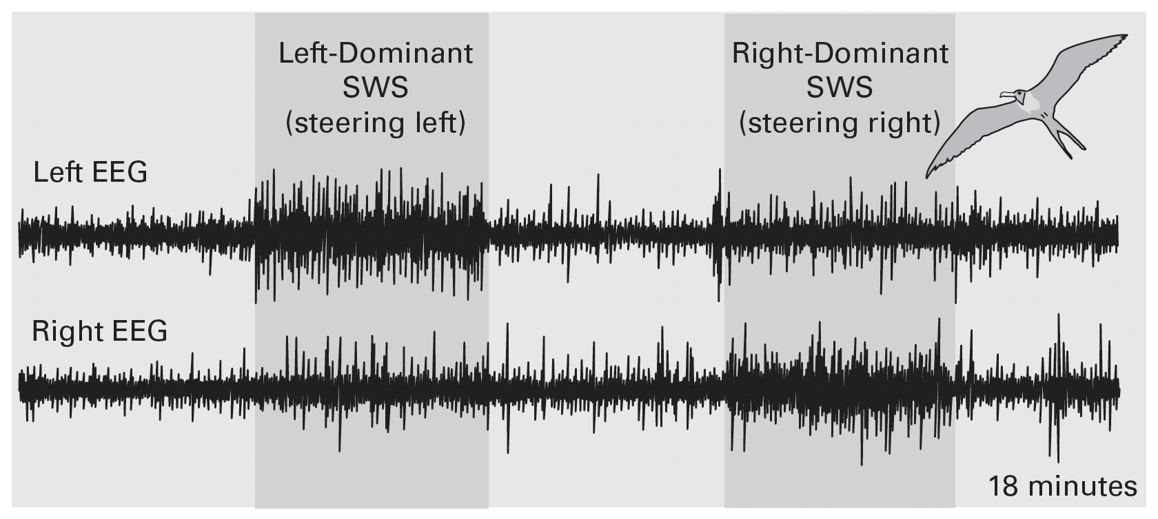
Research image of asymmetric EEG data.
Highly asymmetric sleep in great frigatebirds (figure 2.7): Implanted electrodes and data loggers were used to record the electroencephalogram (EEG) from both the left and right sides of the brain. During this 18-minute clip, the illustrated bird sometimes showed much greater slow-wave sleep (SWS) activity on one side of the brain than the other. Intriguingly, the bird tended to steer left when the left hemisphere “slept more deeply” than the right, and to steer right when SWS was right-side dominant. Adapted from Rattenborg (2017).

Yet another important difference between birds and mammals is that REM sleep in birds is not accompanied by the skeletal muscle paralysis (i.e., atonia) that is typical of mammalian REM sleep. Birds do reduce their muscle tone during REM sleep but not as dramatically as mammals do. In particular, birds during REM sleep maintain significant muscle tone in their legs, which allows them to remain standing while asleep. Ostriches, geese and other long-necked birds also maintain tone in their neck muscles, though their heads may droop or sway a bit during REM sleep. In geese, neck muscle tone is variable: When geese sleep with their head resting on their back, neck muscle tone during REM is lower than it is when the animals are sleeping with their head held high. Apparently, the level of neck muscle atonia during REM sleep is adjustable in birds; no analogous phenomenon has ever been described in mammals.

Sleeping with one hemisphere

M

ost mammals sleep with both halves of their brain at the same time; whenever one cerebral hemisphere is in SWS or REM sleep, so is the other one. The main exceptions are dolphins and whales, which sleep with one hemisphere at a time. This remarkable ability is probably an adaptation to aquatic life, since full-blown sleep, especially REM with its muscle atonia, would impair the ability of a sleeping dolphin or whale to breathe at the surface. In addition, always keeping at least one hemisphere awake helps aquatic mammals remain alert to threats; such vigilance is especially important in open water, where hiding is impossible.

Studies of the EEG in birds have reported several cases of asymmetric slow-wave activity. This activity may not be truly unihemispheric sleep, but the data clearly indicate that these birds sleep more deeply with one hemisphere than the other (figure 2.7). This phenomenon may well be related to the fact that birds and all nonavian reptiles lack a corpus callosum, which interconnects the two cerebral hemispheres in most mammals and likely allows slow-wave activity to spread between them. In this context, it is interesting to note that the corpus callosum is proportionately much smaller in dolphins and whales than in other placental mammals.

As mentioned earlier, some birds regularly sleep with one eye open (and so do some crocodilians). Intriguingly, the open eye is generally opposite the less deeply sleeping hemisphere. The most likely explanation for this phenomenon is that in birds and nonavian reptiles, the visual pathway from the retina to the forebrain is almost completely crossed, which means that visual information from the open eye of an asymmetrically sleeping bird is being sent to the “more awake” hemisphere. This process, in turn, should allow a sleeping bird to remain at least somewhat alert to threats presented to its open eye. In support of this hypothesis, when mallard ducks sleep asymmetrically with one eye open, they tend to direct their open eye (opposite the more awake hemisphere) toward potentially approaching predators. Indeed, these ducks display escape behavior when they see a predator through their one open eye, even while their EEG reveals that one of their two hemispheres is soundly asleep.

During their annual migrations, some birds fly for several days without a break (see Chapter 4), and some of them sleep on the wing. Particularly interesting are frigatebirds, tropical seabirds related to pelicans. EEG recordings from frigatebirds during their long-distance migration revealed that these birds occasionally sleep during flight (although they sleep far more when they do get a chance to land). Some of their in-flight SWS is so asymmetrical that it fits the mammalian criteria for unihemispheric sleep. During these periods, the birds tend to steer left when sleeping more deeply with the left hemisphere; conversely, they steer right when the right hemisphere exhibits more slow-wave activity (figure 2.7). Flying frigatebirds even enter REM sleep, albeit for very brief moments, and during these bouts their head tends to wobble. Thankfully, these moments of in-flight REM sleep are not accompanied by full muscle atonia, which would, of course, cause the birds to fall out of the sky.

How can some birds maintain enough control to fly while drifting in and out of the waking state? Frigatebirds apparently sleep only while they are gliding or soaring, rather than flapping, but how do they maintain sufficient control over their neck, wing and tail muscles to remain afloat? We don’t know the answers to these questions yet, but with further miniaturization of electronics and advances in telemetry, the answers will soon be within reach.

Excerpted from  “Bird Brains and Behavior: A Synthesis” by Andrew Iwaniuk and Georg Striedter. Reprinted with permission from the MIT Press. Copyright 2025. 

Sign up for our weekly newsletter.

Catch up on what you missed from our recent coverage, and get breaking news alerts.