Listen to this story:
0:00
/
Cerebrospinal fluid shows brain-region-specific dynamics, a new high-resolution MRI approach reveals.
A new method captures in rich detail how cerebrospinal fluid swirls in the human brain, and it could help researchers understand the forces that control this complex choreography, according to a new study.
“Everyone is scrambling to find a way to measure [CSF movement in humans], because there’s lots of real reasons to think that it’s an important bodily system and matters for lots of things,” says Douglas Kelley, professor of mechanical engineering at the University of Rochester, who was not involved with the new work. “But it’s really hard to measure directly.”
Most work to date has been done in rodents. Because CSF flows not only through ventricles and the subarachnoid space between the brain and the meninges, but also through tight perivascular spaces that surround small penetrating blood vessels, it is difficult to track via traditional neuroimaging. And even one of the most advanced approaches in humans, intrathecal contrast-enhanced MRI, calls for injecting tracking tracers into the spinal cord, which is invasive, can affect blood flow and thereby muddy the data, and, because the tracers take time to accumulate, lacks temporal resolution.
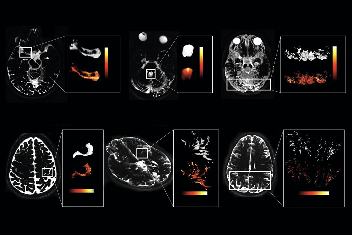
The new method uses high-resolution MRI to provide a noninvasive, granular view of CSF movement—even in the small spaces that surround blood vessels, says study investigator Matthias van Osch, professor of radiology and experimental cerebrovascular imaging at the University of Leiden. “We are the first to actually have the resolution that you actually can measure in these perivascular spaces in the white matter.” The work was published last month in Nature Neuroscience.
The technique could help to expose the diverse range of CSF dynamics at play in the human brain, as well as the forces that drive them, Kelley says, and it pushes “the field closer to being able to make reliable [CSF] measurements in humans.”
T
he new approach, called CSF-STREAM, relies on an ultra-high-resolution 7 tesla MRI instead of invasive tracers or fMRI, which lacks spatial resolution. The CSF-STREAM MRI also features a long echo time—or the interval between the excitation pulse and the peak of the received signal—to suppress signals from other liquids, such as blood, and isolate the CSF signal.CSF moves faster in the subarachnoid space around the large arteries at the base of the brain and ventricles than it does in the tiny perivascular spaces within the basal ganglia and white matter, the team found when they visualized the brains of 20 healthy people.
These dynamics show that CSF flow is facilitated along large blood vessels, says Per Kristian Eide, professor of neurosurgery at University of Oslo, who has conducted intrathecal MRI studies but was not involved in the new work. Eide previously documented compartmentalization within the subarachnoid space, where increased amounts of tracer accumulate, according to a 2024 Nature Communications study. The new work shows a similar pattern of compartmentalization, suggesting that the subarachnoid is “not only an open space,” Eide says.
CSF mobility also fluctuates in tune with cardiac and respiratory cycles, which the researchers tracked in 11 of the participants. But the influence of these driving forces varies by region, too.
At the base of the brain, heart rate affects the pace of CSF movement much more than respiration does, but in the perivascular spaces, cardiac activity and respiration contribute equally. This could be because blood enters the brain through these large arteries but its effect on CSF dampens as it flows into narrower branches, says study investigator Lydiane Hirschler, assistant professor of radiology at the University of Leiden.
Previous lines of evidence suggested that respiration is the main driver of CSF flow, Eide says, but the new study suggests that it “seems to depend on where you are measuring it.”
The findings are in line with previous work showing the role of these factors in driving CSF movement, but they “provide evidence beyond what we had before,” Kelley adds.
One drawback of the new MRI method is that it does not indicate directionality, only how quickly CSF is moving in a specific area of a brain region, Eide points out.
Pinpointing CSF direction will be an important factor in studying the glymphatic system, a proposed mechanism by which the flow of CSF through a network of perivascular spaces removes waste from the brain, van Osch says. Still, he says, their tool could help the field begin to address long-standing questions: “In the end, you want to study the brain cleaning system as close[ly] as possible.”