Drafting a ‘dysfunctome’: Faulty connections to subthalamic nucleus characterize disparate brain disorders

Different circuits between the millimeters-wide structure and the cortex go awry in Tourette syndrome, obsessive-compulsive disorder, dystonia and Parkinson’s disease, a new study of human brain scans suggests.

Making maps: The ‘dysfunctome’ traces circuits that are likely disrupted across different brain disorders.

When Andreas Horn began researching brain disorders treated by deep brain stimulation (DBS) about 10 years ago, he says he also started to wonder about the subthalamic nucleus: How could modulating such a small structure at the base of the brain help people with symptoms as different as tics, tremors and compulsions?

One reason is that targeting that area activates different circuits associated with different conditions, according to a new study led by Horn, associate professor in neurology at Harvard Medical School. He and his colleagues identified the circuits by locating DBS electrodes in brain scans from nearly 200 people with Tourette syndrome, obsessive-compulsive disorder (OCD), Parkinson’s disease or dystonia and plotting those locations onto a normative connectome of the human brain, created using diffusion tractography scans of hundreds of control brains.

The resulting “dysfunctome,” as Horn and his team dubbed it, shows the set of circuits likely to be disrupted in those disorders, Horn says, given that their activation via DBS is associated with symptom improvement.

That’s “not an unreasonable inference,” says Helen Mayberg, professor of neurology and neurosurgery at the Icahn School of Medicine at Mount Sinai, who was not involved in the study. “The subthalamic nucleus is a small area that’s a convergent zone for a lot of different pathways in the brain.”

H

orn’s work figures in an ongoing debate about how best to identify dysfunctional circuitry using diffusion tractography. The method, used to visualize fiber bundles in white matter, provides only an indirect assessment of axonal connections, “but it’s the best we have for noninvasive [measurements of] human brains,” says Sarah Heilbronner, associate professor of neurosurgery at Baylor College of Medicine, who was not involved in the study.

A normative connectome built from tractography scans of control brains provides a low-noise comparison for scans from people with brain disorders, Heilbronner says, but other scientists, Mayberg included, say individualized tractography is needed to capture disease-related differences in anatomical connectivity.

“It’s [something] that we are fighting about relentlessly—there are different camps,” she adds.

Horn’s team purposely used a normative connectome in order to compare across diseases, says study investigator Ningfei Li, a research associate in the neurology department at Charité–Universitätsmedizin Berlin. They aggregated tractography data from 985 people without any brain disorders from the Human Connectome Project to form a connectome of 6 million fiber tracts.

“It’s still a bit controversial, I think, in the field,” Li says, but data from people with brain disorders are hard to come by and often of worse quality than the images included in the Human Connectome Project.

A software program developed by the team, called Lead-DBS, localized electrodes within the subthalamic nucleus on scans from 70 people with dystonia, 94 with Parkinson’s disease, 14 with Tourette syndrome and 19 with OCD treated at seven international DBS centers.
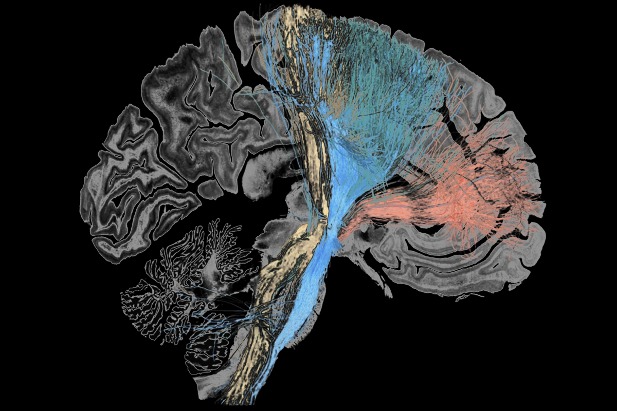
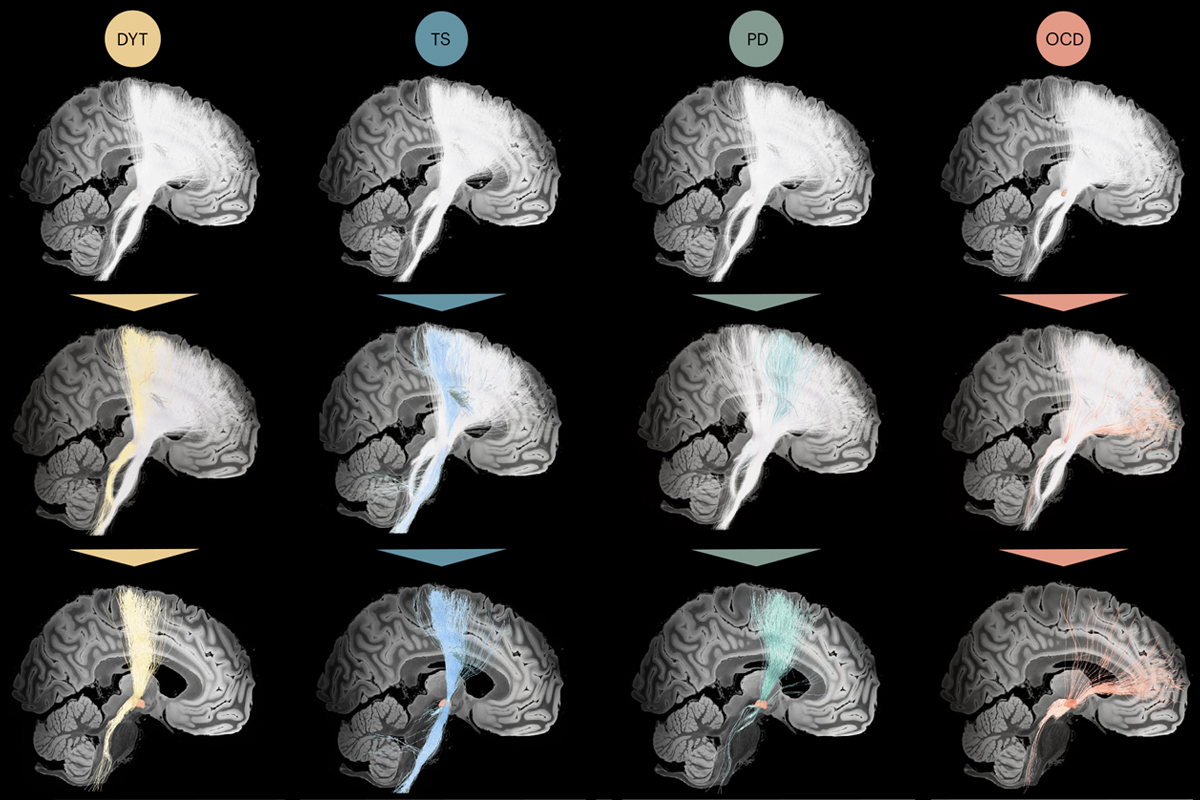
Sweet streamlines: Dystonia (DYT), Tourette syndrome (TS), Parkinson’s disease (PD) and obsessive-compulsive disorder (OCD) are each associated with different circuits between the subthalamic nucleus and the cortex.

Mapping those spots onto the normative connectome revealed “sweet streamlines,” as the team called them. These fibers passed near the electrode sites and, when stimulated, correlated with clinical improvement. Streamlines for Parkinson’s connect to premotor and supplementary motor areas via the subthalamic nucleus, the results suggest, whereas streamlines for Tourette syndrome connect to the primary motor and supplementary motor areas; for dystonia to the somatosensory and primary motor cortices; and for OCD to four areas: the ventromedial prefrontal, dorsal anterior cingulate, dorsolateral prefrontal and orbitofrontal cortices. The findings were published in February in Nature Neuroscience.

T

o better activate the circuits indicated by the streamlines, the team changed the stimulation settings for already implanted electrodes in two people, one with Parkinson’s and one with OCD. Both showed improvement in clinical scores. Symptoms also improved in a third person whose DBS electrode was placed and programmed based on the OCD streamlines.

That said, these streamlines may not be ready for broader clinical deployment, says Cameron McIntyre, professor of biomedical engineering at Duke University. It is challenging to predict which fibers activate in response to electrical stimulation, because of uncertainty about a DBS electrode’s exact location, according to a computational study by McIntyre. “We just don’t have the right tools to be able to dissect which of these pathways are actually related to therapeutic effect,” he says.

There are possible sources of error that would, in theory, preclude his team’s findings, Horn says, “but no matter how we do it, we always show that it’s a more frontal circuit for OCD and a more posterior circuit in dystonia, for example. And in our view, that is a helpful distinction and maybe a contribution.”

Horn says he plans work to refine the new streamlines using more anatomically detailed connectomes, in collaboration with Anastasia Yendiki, associate professor of radiology at Harvard Medical School. Yendiki received about $4.5 million from the BRAIN Initiative in 2023 to image neural circuits from the subthalamic nucleus to the cortex using the latest techniques, including forms of tomography and light-sheet microscopy. These techniques, Yendiki says, can resolve anatomy at the level of individual axons.

The first control human brain is slated to enter the project’s pipeline this year, Yendiki says. If the project is successful, the team plans to move on to brains from those with brain disorders “and figure out if there’s some difference in how the brains are wired,” she says.

Correction:

Details of Yendiki's grant have been updated in the story.