Listen to this story:
0:00
/
The findings could influence how researchers interpret signals from techniques that use blood flow as a surrogate for neuronal activity.
That active neurons boost nearby blood flow is well known—and widely exploited by neuroimaging techniques to estimate neuronal activity. But only a subset of neurons account for those changes in the brain’s blood supply, a new study in mice shows.
The findings, published today in Nature, suggest that neurovascular coupling is not as straightforward as previous models made it seem, says Patrick Drew, professor of engineering science and mechanics, and of neurosurgery, biology and biomedical engineering at Pennsylvania State University, who was not involved in the study. The coupling is not determined by “all the neurons everywhere,” he says. “If you’re just taking all the neurons and adding up their signal, you might get a different view of what’s going on.”
Previous studies of the influence of neuronal activity on blood flow typically used bulk firing rates from neuronal populations, so “how tightly related [they are] had been sort of up in the air,” Drew says.
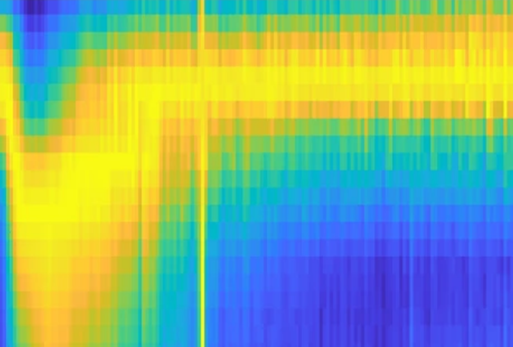
The new study used more granular Neuropixels recordings and functional ultrasound imaging to reveal that two neuron populations account for blood volume changes during arousal, specifically when mice move their whiskers to sense their surroundings. One neuronal population, called arousal-plus, fires more, whereas the other, called arousal-minus, fires less. The activity of these two groups predicted changes in blood volume better than did bulk firing rates across different behavioral states and brain areas, the team found.
If the new findings hold up in people, the fact that blood flow is linked with neurons that are modulated by arousal suggests that functional MRI studies need to take brain state and arousal into consideration, says study investigator Matteo Carandini, professor of visual neuroscience at University College London.
“In the living brain, vasodilation is controlled by neural activity, and it’s a beautiful link,” Carandini says. “It’s just slightly more complicated than people thought.”
B
Arousal-plus neurons fired more during wakefulness and less during sleep, and arousal-minus neurons did the opposite, automated behavioral tracking revealed. These neuronal activity patterns again predicted blood volume changes better than bulk firing did, whether the mice were asleep or awake.
“There’s something really important about sleep and vasodilation and our arousal state. And they seem to be very, very tightly linked,” Drew says.
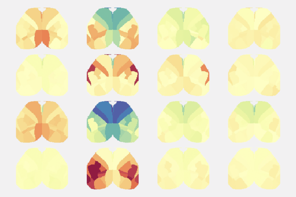
The proportion of arousal-plus and minus neurons varies across the brain, based on recordings across almost 19,000 neurons. For example, arousal-plus neurons are more abundant in the thalamus, whereas arousal-minus neurons are plentiful in the secondary somatosensory cortex. But despite the different proportions, the neurovascular link is global: “It was the same coupling,” Carandini says.
That coupling similarity is surprising because subcortical and cortical regions have different types of neurons, says Alberto Vazquez, research associate professor of neuroscience at the University of Pittsburgh, who was not involved in the work. “We know that the neuronal population is not the same, and, still, similar rules can apply [across these regions],” he says. “Those blood flow regulation characteristics seem to be conserved.”
S
To avoid this potential confounding, mouse studies typically measure blood flow in anesthetized animals, Drew points out. Now, however, behavior-associated blood flow changes prove the need to look at these fluctuations and take arousal into consideration, he says.
Ultimately, the findings complicate the relationship between blood flow and neuronal activity. “The blood signal reflects more than just bulk firing rate that, in a way, holds much more information,” Landemard says.
At the same time, it also makes it trickier to interpret results from neuroimaging technologies, such as blood oxygen level dependent (BOLD) fMRI, that measure blood oxygen as a proxy for neuronal activity, Carandini adds. Studies have already shown that higher BOLD signals, which should reflect higher neuronal activity, actually occur alongside decreased oxygen metabolism.
“It just became much easier to go forward from neural activity to blood and harder to go backwards from blood to neural activity,” Carandini says. “We need, even in our own lab, to deal with the consequences.”