This article is more than five years old.
Neuroscience—and science in general—is constantly evolving, so older articles may contain information or theories that have been reevaluated since their original publication date.
Super-resolution microscopy can set off a series of biological processes that lead to cell death, compromising imaging experiments.
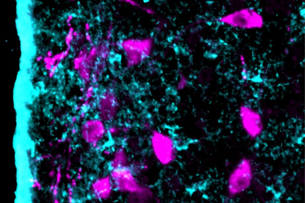
Super-resolution microscopy can illuminate the activities of single molecules within living cells. Scientists have assumed that the method, which uses intense light, is harmless to the cells. But a new study suggests otherwise.
Super-resolution microscopy can instantly kill cells or trigger biological processes that lead to cell death hours later, according to a study published 20 October in Nature Scientific Reports1. The findings suggest that images captured mere minutes into an experiment may reflect cells in distress.
The report outlines methodological tweaks that can help researchers capture healthy cellular processes rather than those of cells about to die.
During super-resolution imaging, researchers label cells with fluorescent dyes or engineer them to express fluorescent proteins, which glow at different wavelengths. The researchers then shine a bright light of a given wavelength onto the cells to illuminate particular ones under the microscope.
The intense light required for super-resolution microscopy can dull the fluorescent dye’s glow after several minutes. It is also known to generate reactive molecules that could harm cells. For these reasons, researchers keep super-resolution imaging experiments short — usually no longer than four minutes. But how cells fare during the experiment and beyond has been unclear.
In the new study, researchers performed super-resolution microscopy on two human cell lines, including immortalized HeLa cells, and one monkey cell line. They first examined the effects of light alone, varying the intensity, wavelength and exposure time. Then they added a fluorescent dye or protein tag to determine whether either one worsens the injury, and monitored the cells for 24 hours.
Cells from all three lines appeared healthy after exposure to intense light at gentle, long red wavelengths for four minutes. But six hours later, some stopped dividing and others initiated the process of programmed cell death, or apoptosis.
At short near-ultraviolet wavelengths, even dim light instantly killed cells in two of the cell lines. Fewer cells died when the researchers replaced the continuous stream of light with a series of rapid flashes. The robust and immortal HeLa cell line survived light that was up to seven times more intense than levels that proved lethal to the other cells.
To determine the effects of fluorescent tags or dyes, the researchers labeled cells from one of the human cell lines with a bright orange dye. They also genetically engineered cells to express fluorescent green or blue proteins. Each treatment triggered apoptosis when the cells were exposed to light, with a combination of both treatments exacerbating the carnage. Adding vitamin C to the cells’ diet protected the cells, suggesting the nutrient helps to prevent light-induced damage.
The findings suggest that using red light and providing a healthy dose of vitamin C could protect cells from the side effects of super-resolution microscopy, allowing researchers to focus on the cellular processes they really want to see.