Glow up: An infant marmoset’s brain and spinal cord (upper left, next to coin for scale; and center, in magnified coronal sections) shows widespread expression of a fluorescent protein after prenatal viral injection.

Prenatal viral injections prime primate brain for study

The approach makes it possible to deploy tools such as CRISPR and optogenetics across the monkey brain before birth.

By Angie Voyles Askham
22 January 2026 | 5 min read

A new method for delivering genes to the primate brain in utero enables researchers to adapt existing genetic tools to monkeys for the first time, according to a new study.

The approach could be particularly useful for studying development, says Cory Miller, professor of psychology at the University of California, San Diego, who was not involved in the work. “It’s super exciting.”

To genetically modify a nonhuman primate, scientists previously had three tools: Create a transgenic animal, which takes years to breed; inject a wildtype animal’s brain with a harmless adeno-associated virus carrying a gene, which remains localized to the injection site; or administer the virus intravenously, which requires large volumes to reach the brain, says study investigator David Leopold, chief of the Systems Neurodevelopment Laboratory at the U.S. National Institute of Mental Health.

The new method adapts a protocol from mice that involves injecting a small amount of AAV into the brain’s cerebrospinal-fluid-filled ventricles. When given to newborn mice in this way, the virus transduces cells throughout the brain with long-lasting results. The approach is less effective in older mice, Leopold says.

The newborn primate brain is more fully developed than the newborn mouse brain. So to get high levels of virus throughout the brain, Leopold and his colleagues hypothesized that they might need to treat monkeys in utero and designed an ultrasound-guided system to do so.

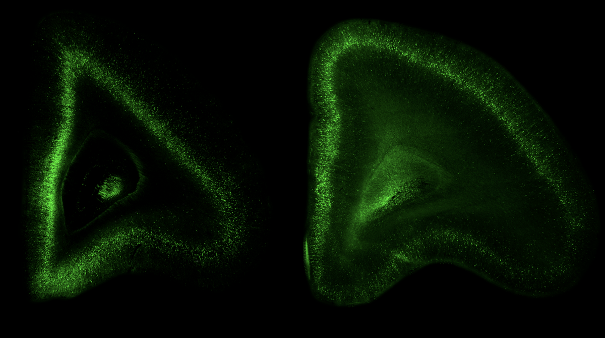
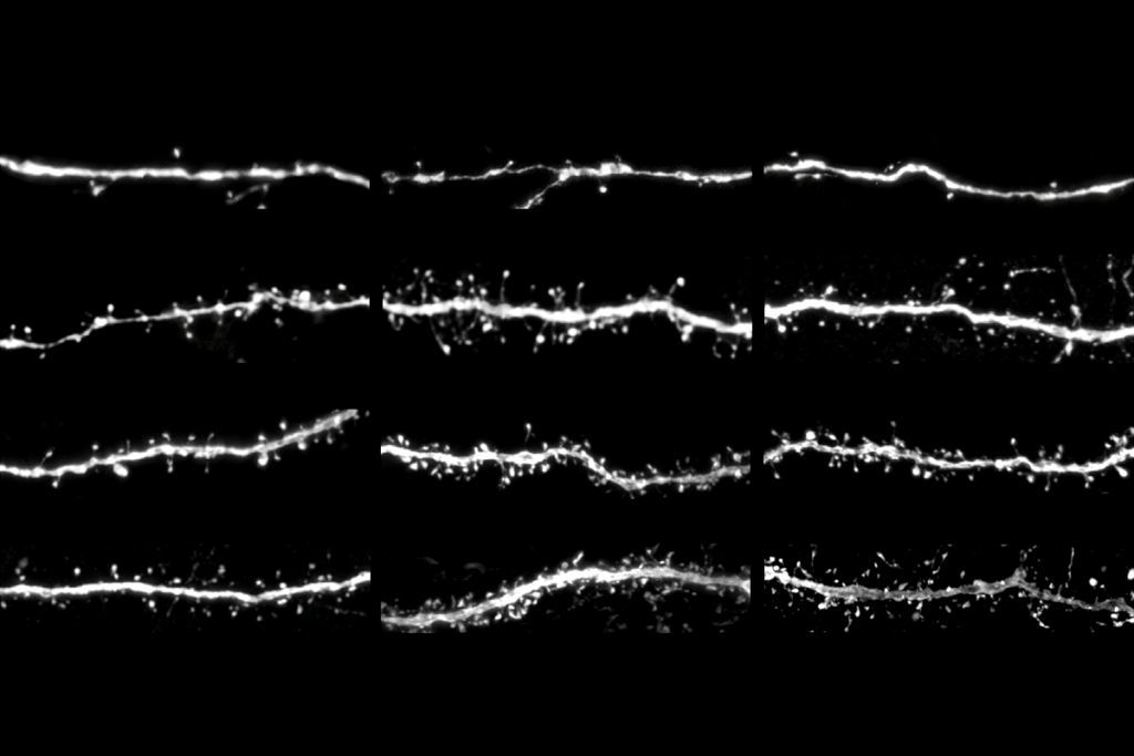
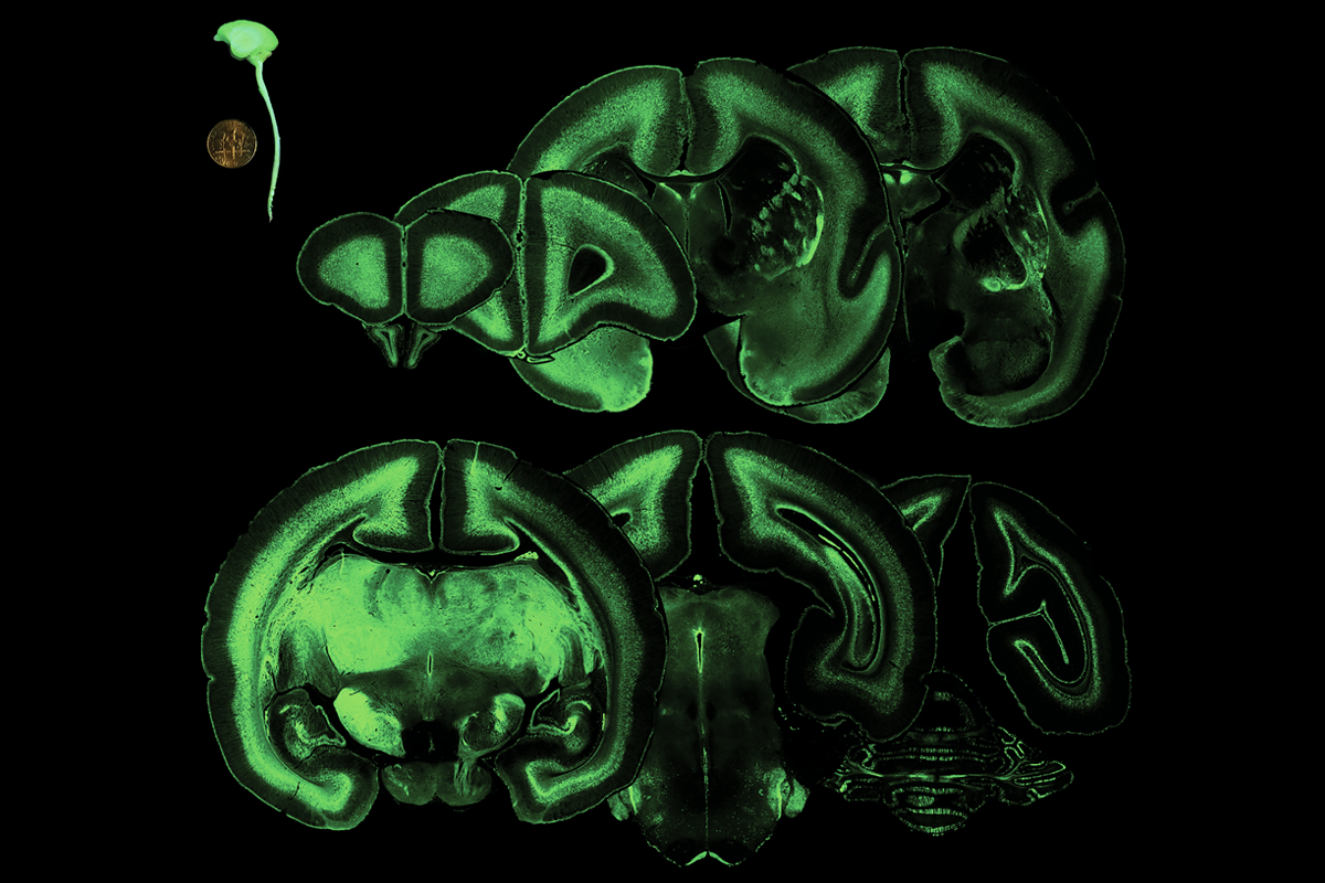
Doing so results in widespread viral expression throughout the animal’s brain that persists for more than two years after injection, the new work shows. The virus can be manipulated to express fluorescent tags or a CRISPR enzyme, and has potential for use with opsins to control cells with light. 

“This work is about actually bringing all the genetic engineering technology into nonhuman primate models, and trying to do this as efficiently as possible,” says Afonso Silva, professor of translational neuroimaging and neurobiology at the University of Pittsburgh, who was not involved in the work.

D

epending on the injection timing and viruses used, the team can label different cell populations, Leopold and his colleagues found. For example, injecting rAAV2 at a particular time in development labels neurons that reside in the late-developing superficial layers of cortex, whereas injecting rAAV9 at that same time marks neurons that reside in deeper layers of cortex, which form earlier.

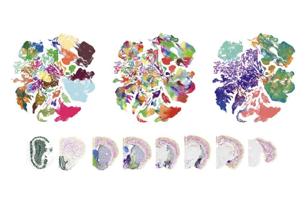
In another application for studying brain development, the researchers time-stamped the birth of a group of neurons by injecting a marmoset with two viruses. One virus produces the Cre enzyme in the presence of nestin, a protein temporarily expressed in newborn neurons. The other carries a red fluorescent reporter that activates when Cre is present. Neurons that appeared red in the animals’ brains had been expressing nestin, and therefore were recently born, at the time of injection.

Time sensitive: Marmosets injected with the same viral delivery system at 75 days (left) versus 94 days (right) post-conception show labeling of different cortical layers.

The intrauterine injection approach also works in rats: The team paired it with CRISPR to introduce a fluorescent tag into the actin gene, which resulted in sparse labeling that is clearly visible in cells’ dendritic spines, where actin is robustly used. 

The method works in the periphery, too, the team demonstrated in rats. A virus injected into the animals’ ventricles before birth labeled cells in the dorsal root ganglia, trigeminal ganglia and peripheral nerves four days later. Injecting a virus into an animal’s eye tagged the axon terminals of retinal ganglion cells, which can reveal these cells’ projection targets within the brain. The study was published last month in Cell Reports.

“This could be a very fast and efficient genetic manipulation tool that can be used across many different species,” says study investigator Kuan Hong Wang, professor of neuroscience at the University of Rochester Medical Center. “It’s kind of a non-transgenic transgenic.” 

T

he new method does require technical expertise and access to specialized equipment, such as an ultrasound machine, Silva says. 

But the injection skills can be learned, and the result is more affordable than many of the alternatives, Miller says. “It’s within the wheelhouse of a lot of labs—a lot more labs than would have the capacity to do full-on transgenic germline work,” he says. Still, current challenges around research funding may make labs hesitant to take on such an ambitious project, Miller adds. “It’ll probably take a few years before it gets more widely adopted.”

Multiple primate labs and centers have already reached out to discuss how they can implement the protocol, Wang says. He says that he and his colleagues plan to use the method to investigate the brain-body connection through the peripheral labeling that the new method enables.

Leopold says he aims to investigate how neuronal projections change throughout development, with the goal of understanding how higher-order visual areas, such as the face patch network, get wired up. “In some ways, the sky’s the limit.”

Sign up for the weekly Spectrum newsletter.

Stay current with the latest advancements in autism research.

privacy consent banner

Privacy Preference

We use cookies to provide you with the best online experience. By clicking “Accept All,” you help us understand how our site is used and enhance its performance. You can change your choice at any time. To learn more, please visit our Privacy Policy.