Spotted: Social cells; brain bulge
A cluster of neurons helps monkeys cooperate, and a human gene makes a mouse brain look like a person’s.
-
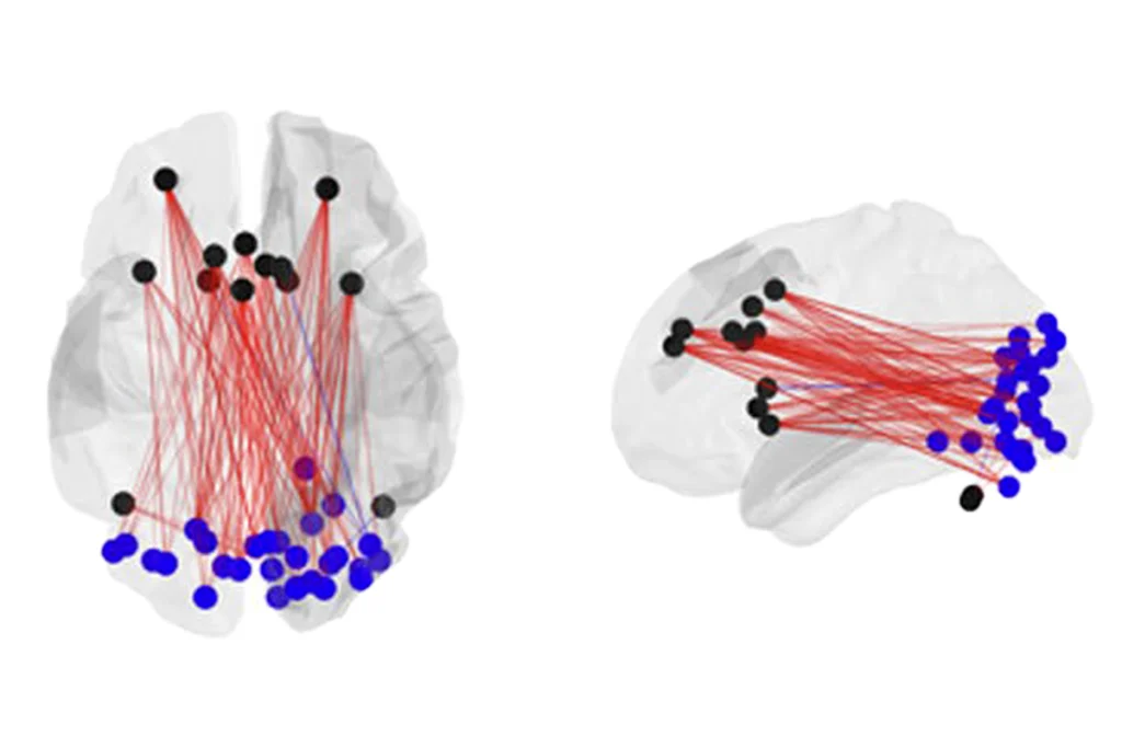
A small cluster of neurons nestled inside the brain helps monkeys cooperate, according to a study published 26 February in Cell. The neurons, which form a C-shaped brain region called the anterior cingulate gyrus, fire more frequently when two monkeys choose to work together toward a common goal than when one monkey puts its own interests first. The neurons even keep track of whether the decision to cooperate was mutual or whether the other monkey ‘defected,’ presumably helping to hone future decisions. The findings could hold clues to autism, as people with the disorder tend to have difficulty anticipating and responding to the actions of others.
-
Autism Speaks turned 10 this week — a big milestone for an organization launched by the grandparents of a child with autism. “When our grandson Christian was diagnosed, there was nowhere for families to turn,” cofounder Suzanne Wright said in a media statement released on 25 February. “By sharing what we’ve learned and taking our message to Washington, the United Nations and even the Vatican, we’ve been able to raise awareness and bring hope to millions of families.” Autism Speaks doled out more than $525 million in its first decade, much of it funding scientific research.
-
Researchers have uncovered previously unknown cell types in the mouse brain, thanks to a technique that measures gene expression in individual cells. Sten Linnarsson, senior researcher in medical biochemistry and biophysics at the Karolinska Institute in Sweden and lead researcher on the study, published 19 February inScience, likened previous efforts to look at gene expression in the brain to running fruit salad through a blender and measuring the color of the juice. The new method, on the other hand, Linnarsson says, “is like taking pieces of the fruit salad, examining them one by one and then sorting them into piles to see how many different kinds of fruit it contains, what they’re made up of and how they interrelate.”
-
Besides being a fraction of the size of our brains, mouse brains are also remarkably smooth — unless they express the human gene ARHGAP11B, that is. In a study published 26 February in Science, injecting the gene into the outer layer of the mouse brain, called the neocortex, induced the proliferation of progenitor cells and caused cortical folding reminiscent of the convoluted surface of the human brain. The findings suggest that ARHGAP11B was involved in the evolutionary expansion of the human neocortex. What mice might do with bigger, bumpier brains, however, remains unclear.
-
Imagine a scanner that can measure brain activity during natural social interactions. The pipe dream has been floating around for a while — it even made the list of ‘Tomorrow’s tools’ for our 2014 Year in Review. A new type of imaging, called diffuse optical tomography, or DOT, brings us one step closer to this dream. The scanner is upright, allowing study participants to sit up rather than lie down, and it’s clear, which means users can talk to other people face to face. Joseph Culver, associate professor of radiology, physics and biomedical engineering at Washington University School of Medicine in St. Louis, is using DOT to study autism. We’ll be keeping an eye on this.
Recommended reading

How pragmatism and passion drive Fred Volkmar—even after retirement

Altered translation in SYNGAP1-deficient mice; and more

CDC autism prevalence numbers warrant attention—but not in the way RFK Jr. proposes
Explore more from The Transmitter
