Progenitors cells in the medial ganglionic eminence become increasingly organized during development as rows of brain imaging progress from top to bottom.
Cellular cradle: The evolutionarily distinct progenitors (red) in the medial ganglionic eminence become increasingly organized during development (top to bottom).
Jia et al, Science.

‘Tour de force’ study flags fount of interneurons in human brain

The newly discovered cell type might point to the origins of the inhibitory imbalance linked to autism and other conditions.

By Holly Barker
29 January 2026 | 4 min read

A new type of progenitor cell found deep in the developing human brain spawns a steady supply of inhibitory interneurons and glia, according to a new study. The findings point to a potential origin of the inhibitory imbalance linked to autism and other conditions. 

The study is a “tour de force” and “adds an important mechanistic piece” to understanding human interneuron diversity, says Xin Jin, associate professor of neuroscience at the Scripps Research Institute, who was not involved in the work.

Compared with other mammals, humans and other primates have larger populations—and a greater diversity—of inhibitory interneurons. It is not completely clear, however, where those extra neurons come from. 

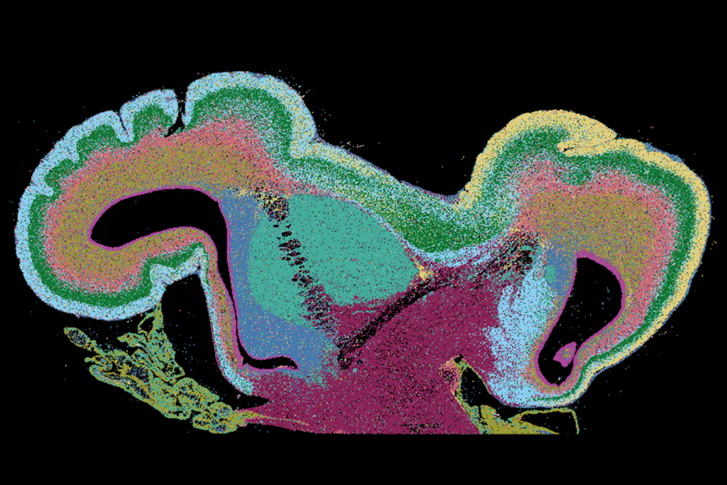
In mice, most interneurons are born in a brain structure called the medial ganglionic eminence and later migrate to the cortex. This structure, which forms transiently during fetal development, might be responsible for the expansion of interneurons during primate evolution, past research suggests. Humans and other primates have an enlarged region within the structure called the subventricular zone, where microglia gather to extend neurogenesis by secreting growth factors. 

That region is also home to progenitor cells that continue to churn out GABAergic inhibitory interneurons into the final stages of pregnancy, the new study found. The cells appear to be “a main driving force for prolonged neurogenesis,” says study investigator Da Mi, associate professor of neuroscience at Tsinghua University. 

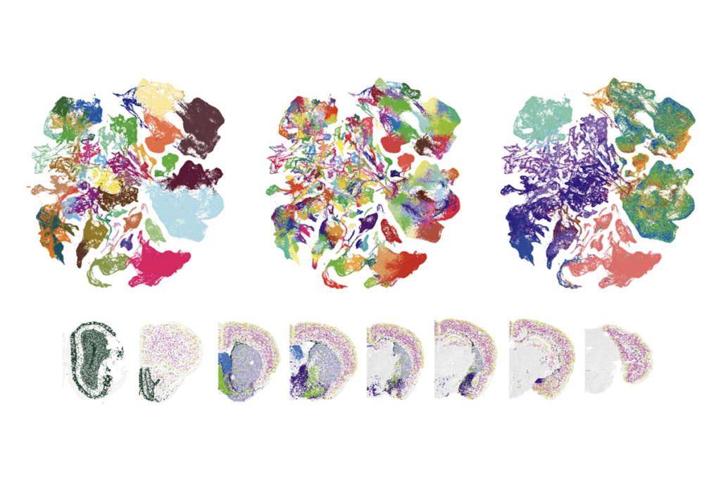
The progenitors—dubbed subventricular zone radial glial cells (SVZ RGCs)—surround nests of proliferative cells that are thought to contribute to human cortical development. Comparing brain tissue samples with those from other mammals, Mi’s team found a similar progenitor population in macaques but not mice, hinting that the cells may have emerged during primate evolution. 

U

sing spatial transcriptomics in human fetal brain tissue from different stages of development revealed seven molecularly distinct clusters of progenitor cells. Each cluster spawns select interneuron subtypes, including interneurons destined for the cortex and striatum, cortical astrocytes and oligodendrocyte precursor cells. 

The findings, published 15 January in Science, challenge previous notions that neuronal progenitors are a homogeneous mass of cells, suggesting that SVZ RGCs activate transcriptional programs that drive cell fate before neurons integrate into circuits. 

“That’s really impactful,” says Tomasz Nowakowski, associate professor of neurological surgery, anatomy and psychiatry, and of behavioral sciences, at the University of California, San Francisco, who was not involved in the new work. “It opens up a lot of questions but potentially provides some molecular handles that we can begin to mechanistically examine,” he adds.

Double take: Dividing SVZ RGCs bear unique characteristics that are observed in other types of progenitor cells.
Jia et al., Science

But linking progenitor clusters to broad classes of interneurons isn’t enough to know their fates, says Gordon Fishell, professor of neurobiology at Harvard Medical School, who was not involved in the work. “They are making a statement about what those cells become, but they are using a few very weak genes to make those conclusions,” he says. “Looking at a radial glial cell is like looking at a pregnant woman and predicting what her child will be like. How would you know?” 

Mi agrees that there are limits to what they know about these cells but counters that mapping the origins of interneuron subtypes would require tracing cells after birth. Interneurons morph into specialized cell types only after they integrate into brain circuits. That kind of longitudinal analysis would be “exceptionally challenging,” Mi says. 

W

hatever the case, SVZ RGCs alone can’t completely account for interneurons’ evolutionary expansion, says Arnold Kriegstein, professor of neurology at the University of California, San Francisco, who was not involved in the study. “It’s more complicated than the appearance of a single [progenitor] cell,” says Kriegstein, whose lab also identified interneuron progenitors in the medial ganglionic eminence of developing fetal brains in a recent preprint.  

For instance, cortical interneurons in the superficial cortical layers—the areas most affected by expansion—are born in the caudal ganglionic eminence, past research has shown. And some interneurons may emerge directly in the cortex, according to a study using human cell culture.  

Mi and his team are now turning their attention to the proliferative nests where SVZ RGCs reside. The group has preliminary data suggesting that the progenitors’ neuronal offspring  drive the formation of these structures, Mi says. But why the nests are more pronounced in humans than in other species, and their potential functions, is something his team hope to uncover, he says.

Sign up for the weekly Spectrum newsletter.

Stay current with the latest advancements in autism research.

privacy consent banner

Privacy Preference

We use cookies to provide you with the best online experience. By clicking “Accept All,” you help us understand how our site is used and enhance its performance. You can change your choice at any time. To learn more, please visit our Privacy Policy.