A diagram of many types of cells
Cell census: The mouse brain has more than 5,000 cell types, categorized here based on their gene-expression patterns.

Welcome to the second single-cell revolution: New high-throughput technologies are transforming how we define neurons

This ongoing essay series will explore questions these technologies raise, as well as opportunities they provide for understanding development, evolution and disease.

Although the brain has been an object of fascination for centuries, neurobiology as we know it originated with Santiago Ramon y Cajal’s magnificent descriptions and drawings of neurons in the late 1800s. His work, which spanned nearly every part of the nervous system of dozens of invertebrate and vertebrate species, set the field’s agenda for the next hundred years: the detailed analysis of single neurons, initially morphologically and later electrophysiologically. It was the first single-cell revolution.

By the end of the 20th century, adherence to this single-cell agenda — coupled with advances in molecular methods, including biochemistry, molecular biology, immunochemistry and transgenesis — had revealed the basics of neuronal structure, function and development. Despite these transformative advances, however, it became increasingly clear that to truly understanding how the brain works — and how it fails in neurological and psychiatric diseases — would require new approaches. To make sense of circuit architecture and pinpoint cells that might be defective in brain diseases, for example, researchers would need to be able to study enough single neurons to classify them into types. That would require the capacity to analyze hundreds to thousands of single cells, preferably simultaneously.

That has finally become possible, with the invention over the past 20 years of a series of massively parallel high-throughput single-cell methods. Today, researchers can molecularly profile cells using high-throughput single-cell and single-nucleus RNA sequencing (scRNA-seq and snRNA-seq). They can monitor neuronal activity with multi-electrode “neuropixel” type probes and genetically encoded calcium and voltage indicators. They can visualize neuronal structure and connectivity with serial-section electron microscopy or optically with super-resolution and expansion microscopy. In each case, they can assay hundreds to thousands of cells in parallel sufficiently quickly and inexpensively (relatively speaking) to classify and characterize neurons and unravel neural circuitry more comprehensively than ever before.

In other words, we are now in the midst of a second single-cell revolution.

This new capacity to accurately classify cell types on a broad scale is revolutionizing how we study neural circuits, providing new insights into brain development and evolution, and opening new avenues for understanding brain diseases. But to fully realize its potential, the field still needs to grapple with a number of questions, such as what the most appropriate level to classify cells is and how closely different facets of cell-type data align. This introductory essay sets the stage for an ongoing series that will examine the uses of this technology for neurobiology, along with challenges that remain.

O

f these new high-throughput methods, none has had a greater impact than sc/snRNA-seq. The method was invented independently and nearly simultaneously by three groups almost 10 years ago — Drop-seq by Evan Macosko and his colleagues, inDrop by Allon Klein and his colleagues, and 10X/GemCode by Grace Zheng and her colleagues. In all three platforms, mRNA from thousands of single cells or nuclei is captured and barcoded, then reverse-transcribed, amplified and sequenced in a single reaction. Cells with similar transcriptomes can be computationally grouped into candidate cell types. Newer methods provide similar results at an even lower cost than the original techniques. By now, researchers have profiled more than a billion single cells.

The first successful effort to use scRNA-seq to generate cell-type atlases of complex tissues used the mouse retina, a particularly accessible part of the brain; we now know that it contains some 130 neuronal types. Researchers have since applied the method to numerous other tissues and species. Over the past few months, Science and Nature have published special issues detailing the largest results from these efforts to date, including expansive atlases of the human and mouse brain. (For more, see “Vast diversity of human brain cell types revealed in trove of new datasets.”)
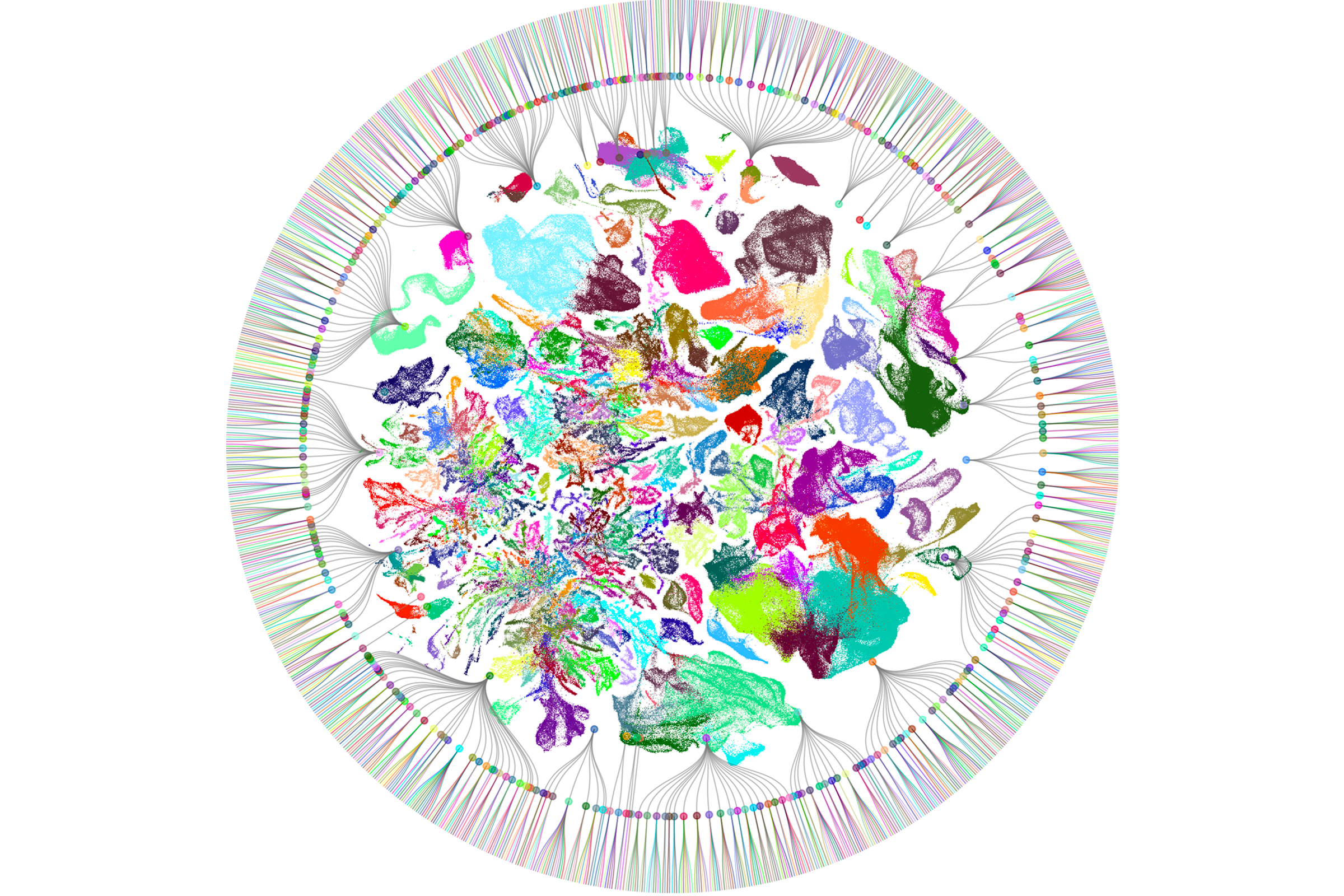
Brain maps: Researchers mapped transcriptionally-defined cells across different regions of the brain.

The atlases, in turn, provide a foundation for addressing many important biological issues: What cell types are affected in neurological and psychiatric diseases? Where are the genes that predispose someone to or cause disease to be expressed? How do cell types that are resilient or vulnerable to insult differ? How do neural cell types diversify, differentiate and mature? How do activity-dependent and activity-independent factors influence these processes? Which cell types are evolutionarily conserved and which arise to meet the needs of particular species?

Sc/snRNA-seq is still relatively new — it has been in wide use for just over five years — so studies have so far provided only partial answers to these questions. To fully address them, the field must overcome several technical and conceptual challenges, set forth below.  Individual essays in this ongoing series will explore some of these specific questions in greater depth, including how cell-type data offer new insight into development and evolution, how to apply different computational methods for grouping cells, and the question of cell type versus state.

        • Localizing cells: Sc/snRNA-seq techniques begin by dissociating cells or nuclei, erasing information on a cell’s location within the tissue. This is unfortunate because neurobiologists routinely rely on location — for example, to map connectivity between brain areas or target cells for recording. New “spatial transcriptomic” methods provide gene expression profiles of cells within tissues, using either multiplexed in situ hybridization to query a select set of genes (e.g., MERFISH) or RNA capture followed by sequencing (e.g., STARmap). Although these methods detect fewer genes than scRNA-seq does, they have become an indispensable adjunct to tissue profiling.


        • Harmonizing criteria: Many researchers viewed the first scRNA-seq-derived atlases with skepticism because it was unclear whether cell types defined by molecular criteria corresponded to those that neurobiologists care about — structure, function and connectivity. In the retina, these different aspects of cell types align very well. But questions remain about other brain regions. Methods that record a single neuron’s physiology, morphology and gene expression will help close this gap. Many to date are relatively low throughput, but combining calcium imaging with spatial transcriptomics holds great promise.


        • Multiomics: The transcriptome has proven useful for classifying cells but can’t yet fully characterize them. RNA levels are imperfectly correlated with protein levels and do not reliably distinguish among alternatively spliced isoforms. Moreover, they fail entirely to identify post-translational modifications, such as glycosylation, phosphorylation and many others, all of which are essential for neuronal function. This shortcoming has led to great interest in “multiomics,” in which RNA-seq is combined with other methods. Researchers have successfully combined RNA-seq and ATAC-seq, which assays chromatin accessibility, a key epigenomic measure. But single-cell proteomic assays have not been optimized or widely used, particularly in combination with other methods.


        • Granularity, types and states: Researchers have not yet settled on the optimal resolution for grouping cells. At one extreme, there could be as many neuronal types as there are neurons. At the other, there could be very few types — for example, only sensory neurons, interneurons and projection neurons. Where is the sweet spot in between? This problem remains unsolved, with multiple computational models proposed to group cells into types, types into classes and so on. Perhaps more troubling is that initial transcriptome-based definitions of cell types made the implicit assumption that once animals reach adulthood, their transcriptomes are stable. Of course this is not true; neurons express different genes at different times, with major changes that depend on activity levels and patterns, hormones and more. Injury or disease lead to even greater changes. It remains challenging to distinguish these differences in cell state — meaning cells of the same type expressing different genes under different conditions — from differences in cell type.


Get alerts for “Defining cell types” in your inbox.

This series explores how new high-throughput technologies are changing the way we define brain cell types — and the challenges that remain.