This article is more than five years old.
Neuroscience—and science in general—is constantly evolving, so older articles may contain information or theories that have been reevaluated since their original publication date.
We asked autism researchers to enter the Spectrum science image contest. Here are the top pics.
We asked autism researchers to enter the Spectrum science image contest. From sensational stem-cell snapshots to a ‘furry’ close-up of the hippocampus, these are the 10 top pics.
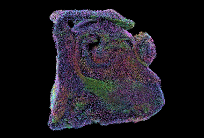

Fuzzy memories: A close-up of a human hippocampus, created by magnetic resonance imaging, shows the short-range connections critical for forming new memories.
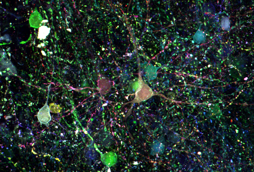
Brainbow brilliance: Individual cells in the human brain reflect one of more than 100 hues after researchers treat the tissue with a combination of four dyes — a technique called ‘Brainbow.’
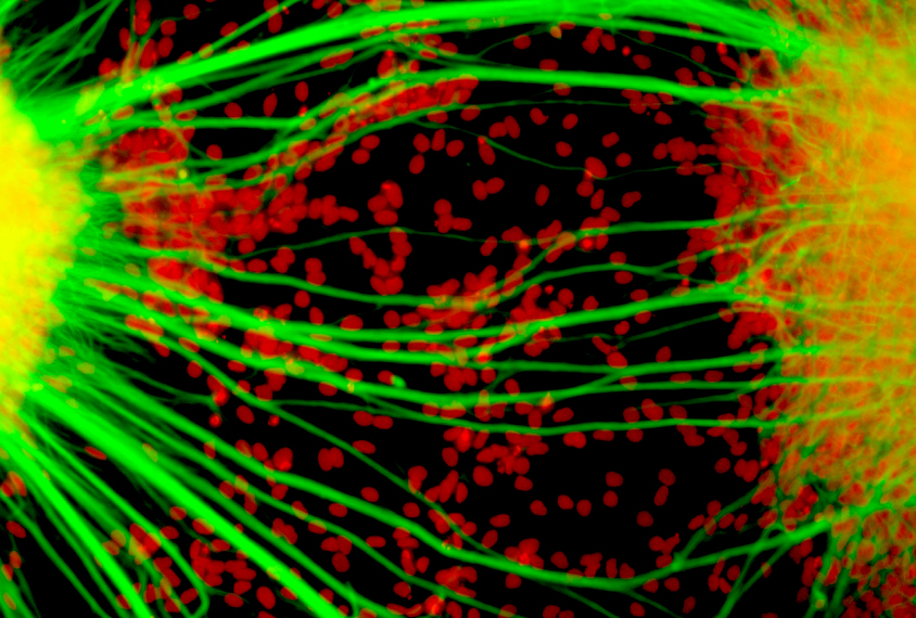
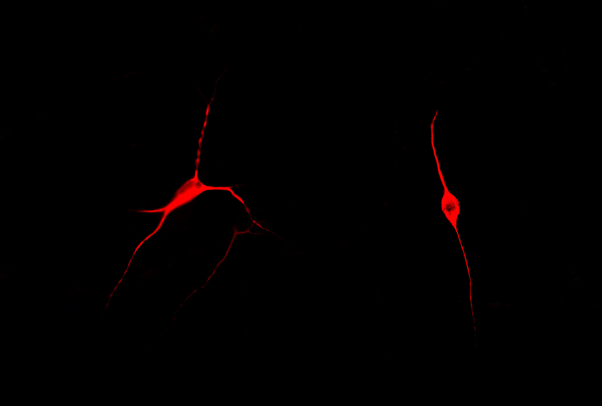
Creating connections: Two clusters of immature neurons (nuclei, red) derived from the cells of a child with autism form a bridge of fibers between them.
Magical mind: Data from magnetic resonance images of multiple human brains blend to form a colorful depiction of the bundles of nerve fibers (right) that transmit information in the brain.
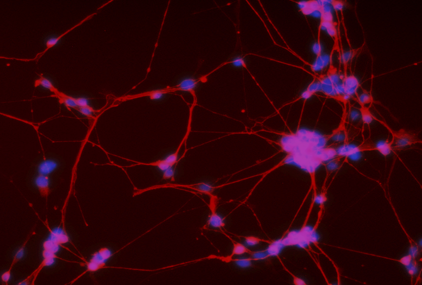
In the pink: After seven days in culture, neural stem cells (pink) derived from a person with autism express a protein called beta-III tubulin (red) present in immature neurons.
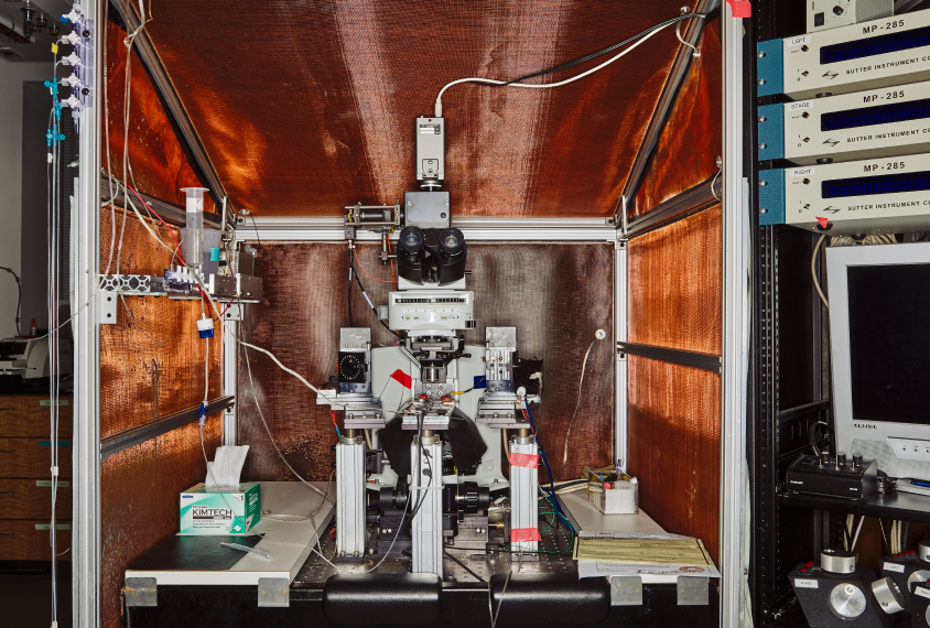
Decoding device: This cell-recording machine captures the electrical activity of specific cells in slices of brain tissue that have been activated by light.
Simple cells: Neurons derived from people with autism have few branches (right). Exposing them to support cells called astrocytes (black) from controls leads to a more typical shape (left).
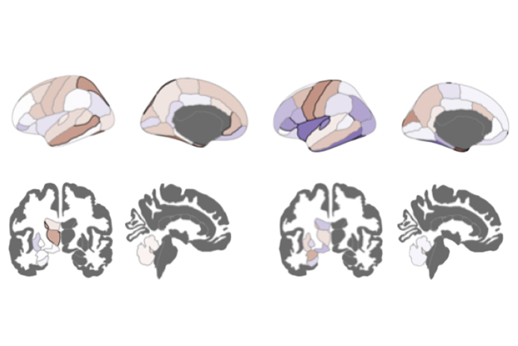
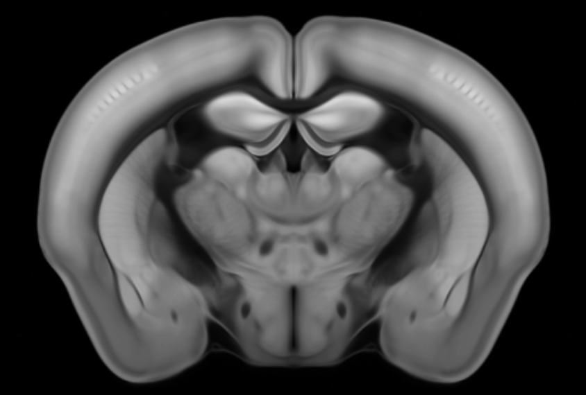
Rodent reveal: A 3-D model of a mouse brain shows more than 500 brain regions in great detail.
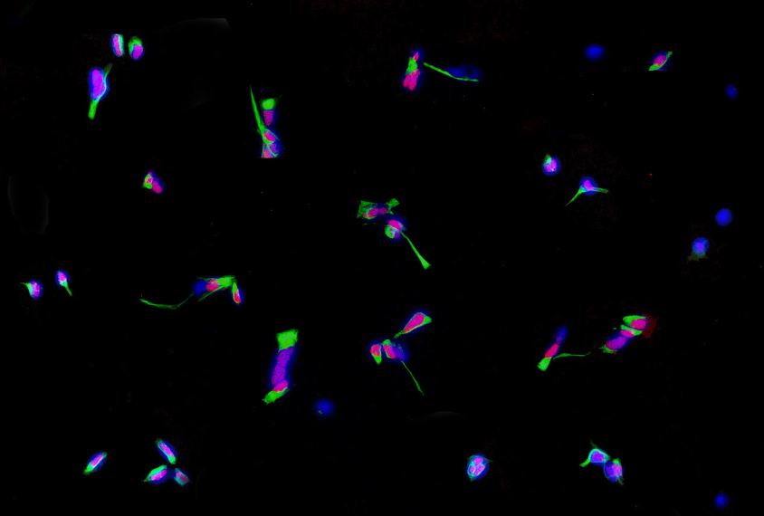
Painted cells: Three separate stains (green, red, blue) highlight precursors of human neurons grown in culture.
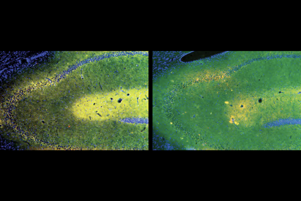
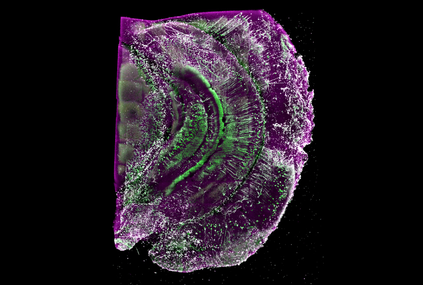
Rhapsody in green: Neurons infected with the rabies virus glow green in the hippocampus (memory hub) of the mouse brain.