Brain scans may forecast effectiveness of autism treatment
Patterns of activity in certain brain regions may predict how well a child with autism will respond to a behavioral therapy.
Patterns of activity in the social brain predict how much a child’s autism features will improve after a behavioral therapy called pivotal response treatment, according to a new study1.
The study is small, but the findings hint that brain scans, or an equivalent technology, could help clinicians select the most promising treatment for a child with autism.
“It takes time and money to try a treatment,” says lead investigator Daniel Yang, assistant research professor of pediatrics at George Washington University in Washington, D.C. “We don’t want to waste time exploring a false hope.”
Clinicians have no proven method for recommending a particular treatment for a child with autism. “But we often find that kids who have similar clinical profiles respond very differently” to a given therapy, says Shafali Jeste, associate professor of psychiatry and neurology at the University of California, Los Angeles, who was not involved in the study.
This variability can complicate tests of autism treatments. A therapy that works in some children may appear ineffective in a larger, more heterogeneous group. A biomarker that predicts treatment response in children with autism may help researchers choose who to enroll in a trial. “That’s why this work is exciting,” Jeste says. The findings appeared in November in Translational Psychiatry.
Functional forecast:
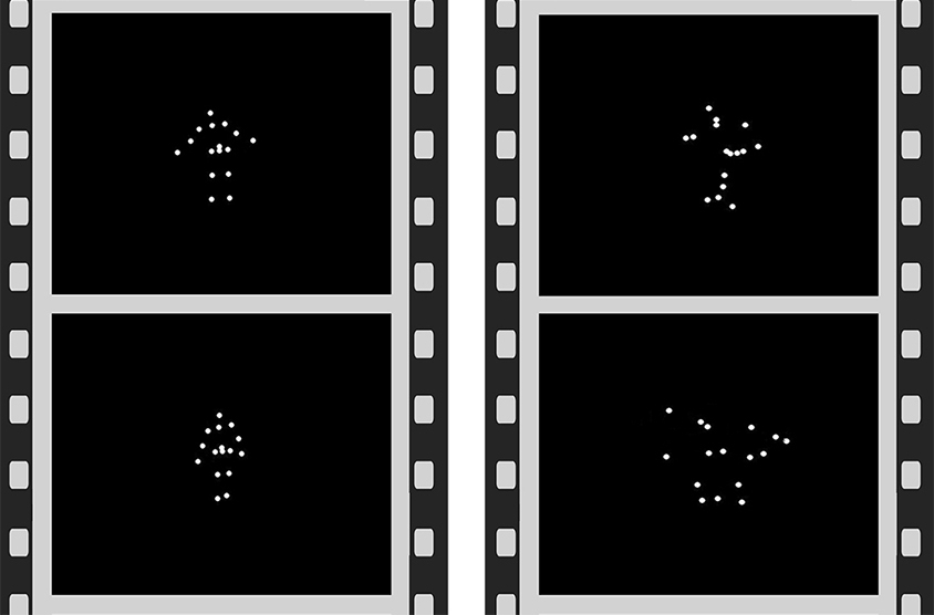
Yang and his colleagues used functional magnetic resonance imaging to record brain activity in 20 children with autism, aged 4 to 7 years, as they watched animations of dots. The dots either moved randomly or resembled a person doing something social, such as playing ‘pat-a-cake.’ The researchers compared brain activity in response to human motion versus random motion.
The children then received seven hours of pivotal response treatment each week for 16 weeks. The therapy involves using activities and objects that interest a child to foster communication and other social behaviors, such as taking turns during play.
The researchers assessed each child before and after treatment using the Social Responsiveness Scale, a parent questionnaire. They looked to see how changes in autism severity track with brain activity prior to treatment.
A statistical analysis identified four clusters of brain regions whose response to the animations predicted improvement after treatment. The more active these regions are in response to human motion relative to random motion, the more effective the treatment is in a child.
All of the clusters are involved in social behavior. One includes the fusiform gyrus and superior temporal sulcus, areas involved in social perception. Another comprises regions on the right side of the brain that govern social attention. A third includes regions that regulate emotion. The fourth encompasses areas implicated in social memory and motivation, including the hippocampus and the amygdala.
Possible predictor:
The researchers then looked to see whether brain activity patterns in these clusters predict treatment effectiveness. They used a machine-learning algorithm to analyze data from 19 of the participants to predict the remaining individual’s response to treatment, and repeated the process for each participant.
The computer predicted 72 percent of the variation in response to treatment.
The findings suggest that clinicians can one day use brain activity patterns to identify children with autism who would benefit from a treatment, Yang says. But wide use of such an approach might depend on clinicians’ ability to gather the data using inexpensive imaging methods or a surrogate technique such as eye tracking.
It is also unclear whether the findings apply to all individuals with autism. The children in the study are not cognitively impaired and can lie motionless in a scanner. These are “definitely not your average kids with autism,” says Antonio Hardan, professor of psychiatry and behavioral sciences at Stanford University in California, who was not involved in the study.
Hardan says the next step is to validate the findings using different methods for measuring autism severity and include a control group of children who do not receive therapy. He is planning to collaborate with the Yang team.
References:
- Yang D. et al. Transl. Psychiatry 6, e948 (2016) PubMed
Recommended reading

New organoid atlas unveils four neurodevelopmental signatures
Explore more from The Transmitter

The Transmitter’s most-read neuroscience book excerpts of 2025

Neuroscience’s leaders, legacies and rising stars of 2025


