The connectivity theory of autism, explained
A growing body of evidence suggests that autism involves atypical communication between brain regions, but how and where in the brain this plays out is unclear.
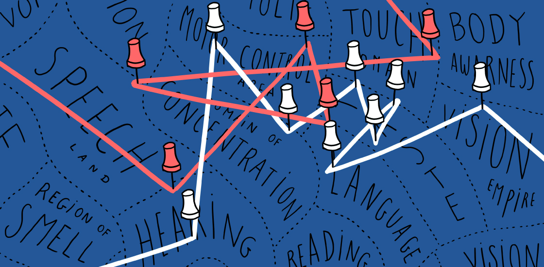
On the face of it, the ‘connectivity theory’ of autism is simple: In autism brains, the theory goes, communication between regions is atypical. But exactly how is complex — and unclear.
By connectivity, researchers generally mean ‘functional connectivity’ — a measure of the degree of synchronized activity between different brain regions. Over the past decade or so, a growing number of autism studies have examined connectivity patterns while participants rest in a brain scanner.
Some of this work indicates that autism is characterized by underconnectivity between distant brain regions and overconnectivity between neighboring ones; others show differences in connectivity within certain brain networks. In one study, connections within the default mode, or ‘daydreaming,’ network of autism brains looked especially weak.
Out of these disparate findings, researchers have constructed the broad-strokes connectivity theory. But the results of some studies are at odds with the proposed patterns, and some find no differences between autism and control brains at all. As studies become bigger and more sophisticated, the number of divergent findings grows.
Here we explain these conflicting results and how researchers are addressing them.
Why are connectivity findings in autism so inconsistent?
One of the main reasons is autism’s diversity: Each person with autism is different from the next, with a unique constellation of behaviors, experiences and genetics. It’s no wonder individual connectivity patterns also differ.
So some researchers have started to look for connectivity patterns in subgroups of people with the condition. For example, mutations in the autism-linked genes MET and CNTNAP2 produce patterns that differ from those in people without these mutations. Connectivity may also differ between men and women with autism.
Large datasets, including the Autism Brain Imaging Data Exchange, may help researchers pinpoint clusters of autistic individuals with distinct connectivity patterns. Some researchers have started searching for clusters on a smaller scale, with a focus on regions of the brain that govern language, for example.
What other factors affect connectivity patterns in the brain?
Age. Several reports suggest that connectivity differs between children and adults with autism. For example, autistic children may have unusually strong connections in several brain networks; autistic adults tend to show weaker connections in some of the same networks.
Homing in on puberty might reveal why their brains appear to switch in this way.
Researchers could also gain insight from studying autistic toddlers before they receive treatment. A study of sleeping 2- to 4-year-olds suggests that connections between some brain regions are unusually weak, not strong, in young children with autism.
The most valuable data come from studies that track connectivity in the same people over time. One of the few studies of this type suggests that connectivity between some brain networks increases from early to late adolescence in typical people but remains stable in autistic individuals. Several studies following children through adolescence are likely to provide more information.
Can research methods influence connectivity results?
Yes. Results from brain imaging studies are subject to variations in magnet strength, experimental approach and methods of data analysis. Connectivity between some brain regions is stronger, for example, when participants’ eyes are open than when they are closed. Psychotropic drugs, such as fluoxetine (Prozac) or methylphenidate (Ritalin), can also alter patterns.
Head movement and other motion, such as breathing, can distort results as well. Researchers are still ironing out ways to minimize or correct for these factors.
Using various imaging techniques in combination may provide richer data and more reliable findings, some say. The most common method is functional magnetic resonance imaging. Other methods include magnetoencephalography, which tracks fluctuations in the brain’s magnetic fields, and diffusion tensor imaging, which maps structural connections.
Could connectivity serve as a biomarker for autism?
It’s unlikely. Some scientists are searching for biological markers that reliably signal the condition, but it has been hard to show consistent differences in connectivity. Patterns of connectivity in autism also may overlap with those of other conditions, such as schizophrenia or depression.
Part of the problem lies in researchers’ uncertainty about how to analyze scans. A method called machine learning is often used to classify participants as autistic based on their connectivity, but different algorithms can produce discrepant findings. And researchers frequently cannot replicate their findings when they apply their methods to a new set of data.
Recommended reading

INSAR takes ‘intentional break’ from annual summer webinar series
Dosage of X or Y chromosome relates to distinct outcomes; and more
Explore more from The Transmitter
Xiao-Jing Wang outlines the future of theoretical neuroscience

Memory study sparks debate over statistical methods
