Painter’s palette: Inhibitory interneurons in the mouse telencephalon are grouped according to cell type (left), subtype (middle and below) and region (right).
Van Velthoven et al., Nature

Constellation of studies charts brain development, offers ‘dramatic revision’

The atlases could pinpoint pathways that determine the fate of cells linked to neurodevelopmental conditions.

By Holly Barker
5 November 2025 | 6 min read

Multiple mouse and human brain atlases track the emergence of distinct cell types during development and uncover some of the pathways that decide a cell’s fate. The findings were published today in a collection of Nature papers.

The papers highlight the timing and location of cell diversification and offer fresh insights into the evolution of those cells. Neuronal subtypes emerge at starkly different times in distinct brain regions, according to multiple mouse studies. And the work upends ideas about cell migration, including the notion that a portion of cortical neurons are made on site, developmental maps of the human brain suggest. 

“This is a dramatic revision of the fundamental principles that we thought were true in the cerebral cortex,” says Tomasz Nowakowski, associate professor of neurological surgery, anatomy and psychiatry, and of behavioral sciences, at the University of California, San Francisco and an investigator on one of the new studies.

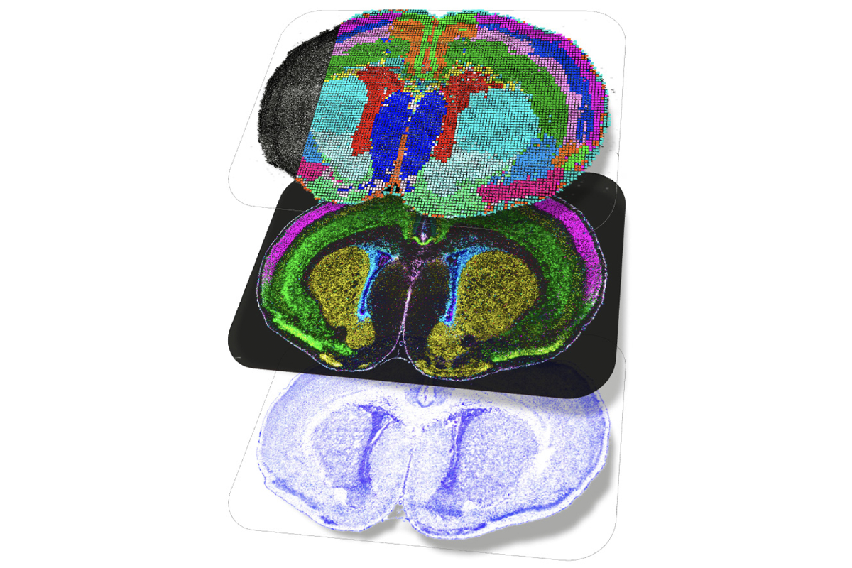
The special issue comprises 12 papers—including 6 newly published ones—from groups working as part of the BRAIN Initiative Cell Atlas Network. The work builds on the network’s complete cell census, published in 2023, that cataloged 34 classes and 5,322 unique cell types in the adult mouse brain.

“Those cell types don’t appear out of a vacuum at the same time,” says Nowakowski, who co-authored a commentary on the new collection. Pinpointing when those cells emerge and where they originate from was the “obvious next question,” he says.

A

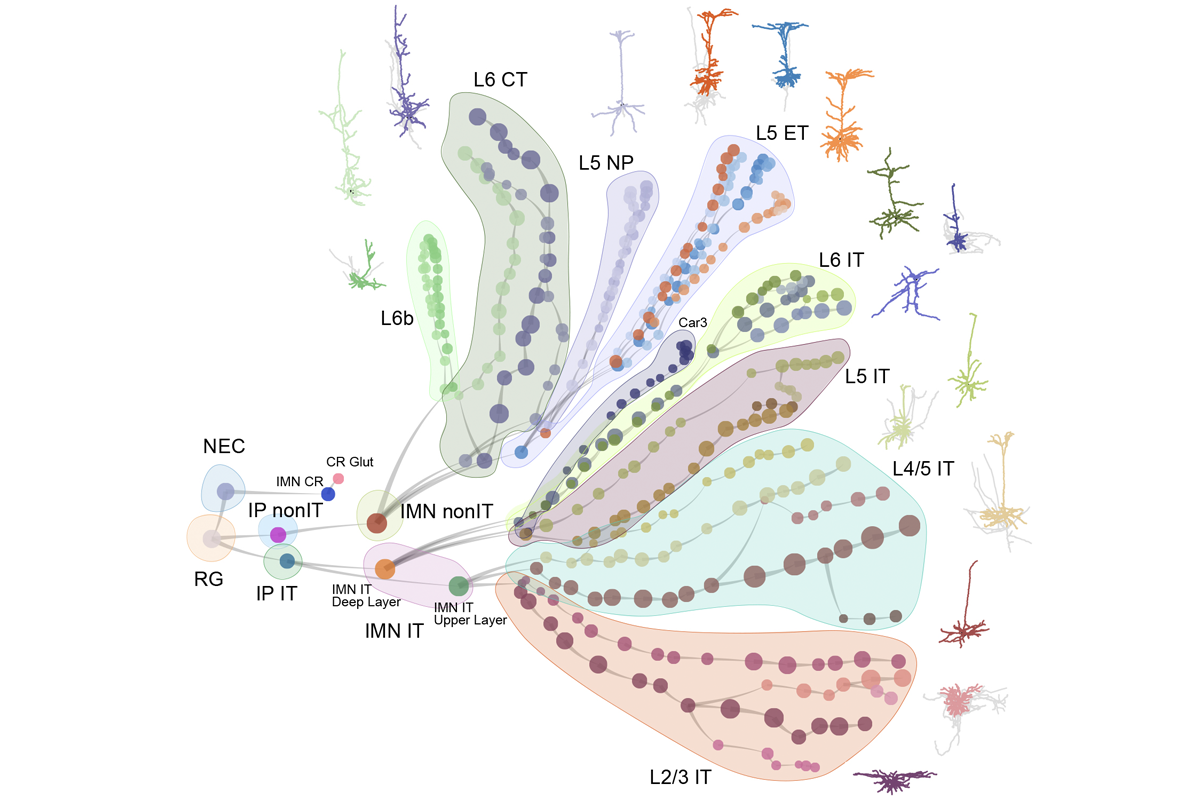
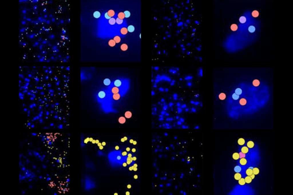
t birth, the mouse brain contains all the initial cell classes that diversify into the multitude of neurons and glia found in older rodents. But precisely when that diversification occurs varies among brain regions: In the visual cortex, new cell types emerge weeks after birth and peak twice—once when the animal first opens its eyes and then again at the onset of the critical period, according to one study.

The findings suggest that in the cortex, a cell’s fate is driven by an interaction of hardwired genetic programs and sensory experience, says study investigator Hongkui Zeng, director of the Allen Institute for Brain Science.

Outside of the cortex, however, diversification mostly takes place before birth, according to a second mouse study from Zeng’s lab. In that paper, Zeng and her colleagues charted the emergence of GABAergic interneurons—a highly diverse group of cells linked to various neurological conditions—in the mouse telencephalon.

Technical triptych: A new map of the mouse brain harnesses three spatial sequencing tools, including transcriptomic, epigenomic and proteomic analysis. The study is one of 12 that together map early brain development across multiple species.
Zhang et al., Nature

Whereas cortical interneurons spawn new cell lineages postnatally, interneurons in the septum and hypothalamus diversify during embryonic development, the study found. In some parts of the hypothalamus, diversification quickly follows neurogenesis, according to another mouse study in the collection.

The delayed diversification in the cortex may extend the period in which the environment can shape neural circuits, says Gregg Wildenberg, a research scientist at the University of Chicago, who was not involved in the studies.

O

ther studies in the package note some key species-specific differences in how subtypes of brain cells emerge. For example, a type of interneuron characterized by its expression of the neuropeptide tachykinin-3 (TAC3) makes up almost one-third of the inhibitory cells in the human striatum, but those interneurons are absent in mice. How humans and other primates evolved a new type of neuron was unclear, says Fenna Krienen, assistant professor of neuroscience at Princeton University.

Cellular destiny: Two transcription factors drive differentiation of deep-layer cortical neurons in cerebral organoids.
Nano et al., Nature Neuroscience

In another new study, Krienen and her collaborators tracked the emergence of interneuron subtypes in the brains of 12 mammalian species, including mice, pigs, macaques and humans. All the mammals share the same initial classes of inhibitory cells—including the group that generates TAC3-expressing interneurons, the study found. In early development, mice suppress the protein and amp up expression of another protein, tyrosine hydroxylase (Th).

The interneurons are known to populate the mouse striatum, but scientists had no idea that they were the murine equivalent of TAC3 interneurons, Krienen says. “They were hiding in plain sight.”

The findings suggest that during evolution, a conserved set of initial classes were tweaked to create new neuronal subtypes, says study investigator Alex Pollen, assistant professor of neurobiology at the University of California, San Francisco. “Evolution acts on neurons not by creating new cell types, but by teaching old neurons new tricks.”

And the source of inhibitory neurons may differ across species. In mice, excitatory neurons are born from progenitor cells in the cortex, whereas inhibitory interneurons originate from progenitors in the ganglionic eminences and then migrate into the cortex. In humans, however, the same cortical progenitor cells produce a local supply of excitatory and inhibitory cells, according to a 2021 paper from Nowakowski’s lab.

Branch out: Excitatory neurons in the mouse visual cortex diversify from a common progenitor into numerous subtypes.
Gao et al., Nature

Rather than producing both cells simultaneously, human cortical progenitors swap production of excitatory neurons for inhibitory cells by the middle of the second trimester, according to a new study from Nowakowski’s team. And a subgroup of progenitors called truncated radial glia keep churning out excitatory neurons throughout the second trimester, at which point mouse progenitors have replaced neurogenesis with gliogenesis, the study also found.

“The value of unbiased atlas studies is that, in addition to answering long-standing questions, they point toward observations that we didn’t know existed and reveal new questions,” Nowakowski says.

M

erging multiple atlases of the developing brain can reveal insights that aren’t apparent from studying a single map alone, another paper in the collection demonstrates. A meta-atlas that combines data generated from 23 human brain atlases from development to adulthood pinpointed gene networks that drive the emergence of neuronal subtypes. One of those networks was amplified in a type of deep-layer cortical neuron that expresses a protein called FEZF2, the study found. Probing the network in a cerebral organoid revealed that FEZF2 interacts with the autism-linked gene TSHZ3 to drive cell fate.

That type of systematic approach could help researchers artificially recreate neuronal subtypes as potential cell therapies for neurodevelopmental conditions, Nowakowski says. “We’re no longer witches at the cauldron, [hoping] to discover small molecules that happen to guide differentiation,” he says. Instead, “we’re using a data-driven approach to derive something meaningful.”

Sign up for the weekly Spectrum newsletter.

Stay current with the latest advancements in autism research.

privacy consent banner

Privacy Preference

We use cookies to provide you with the best online experience. By clicking “Accept All,” you help us understand how our site is used and enhance its performance. You can change your choice at any time. To learn more, please visit our Privacy Policy.