Internal recordings of human brain may offer insight into autism
A technique called intracranial electroencephalography can reveal brain functions with great sensitivity and may ultimately unearth the underpinnings of autism.
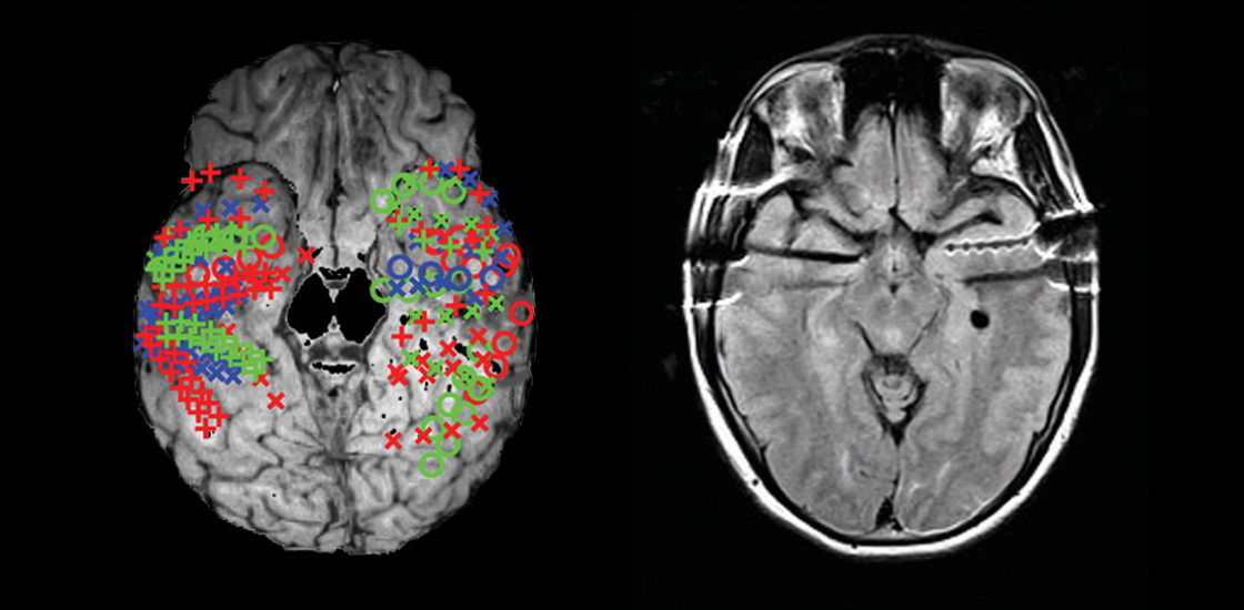
Neuroscientists have an array of tools for understanding the brain. One of the most impressive approaches involves implanting electrodes in the human brain to record neuron activity. It is invasive but is sometimes necessary for medical reasons.
In severe cases of epilepsy, medications don’t work and surgeons try to stop the seizures by removing the region of the brain in which the seizures originate. To find the right spots to remove, doctors implant electrodes into several brain regions and look for bouts of irregular activity.
This approach, called intracranial electroencephalography, or iEEG, is gaining popularity. Numerous research groups now collaborate with hospitals to record iEEG for research purposes.
The technique has much better spatial resolution than traditional electroencephalography (EEG); in EEG, scalp electrodes monitor the summed activity of millions of neurons through the skull.
The iEEG signal measures responses over milliseconds, compared with seconds for functional magnetic resonance imaging (fMRI). Although it typically reflects the activity of hundreds of thousands of neurons, this can be broken down into signals of different frequencies, each of which represents a channel of information flow in the brain. Researchers can analyze iEEG signals recorded at different locations to deduce how parts of the brain interact.
In short, iEEG provides a powerful window into how the brain processes information1. And because epilepsy and autism often co-occur, we have the opportunity to use iEEG to explore how brain processes are altered in autism.
Face value:
The surgery to implant the electrodes for iEEG can last several hours. The number of electrodes and their placement varies. In some cases, the electrodes are placed deep in the brain and in others, along its surface. Once they are implanted, they are hooked up to cables for the recordings. People often spend several days or even weeks in their hospital room, waiting to have a seizure so the electrodes can capture where that seizure originated.
In most cases, the technique picks up electrical signals called local field potentials from a group of neurons. But specialized electrodes can capture signals from single neurons, too. The results have been intriguing2.
For instance, we now know there are neurons that respond specifically to faces — and even to specific faces: In a 2005 study, researchers used iEEG to identify neurons that fire only when a person sees the face of actress Jennifer Aniston3. Some of these neurons seem to track our impressions of a face, such as whether it shows a certain emotion4.
It is also often possible to pass an electrical current through the electrodes in the opposite direction: Rather than record activity, one can disrupt or stimulate a region. This has allowed researchers to influence the perception of a face, as well as to elicit complex mental phenomena, such as a strong sense of willpower5.
Tapping emotion:
One brain region that has received much attention in iEEG studies is the amygdala. This also turns out to be a structure of interest to autism researchers: Amygdala neurons look and behave atypically in autism. Neuroimaging and postmortem brain tissue studies reveal structural abnormalities in the amygdala, and several studies suggest that atypical amygdala activation accompanies autism. The amygdala plays an important role in emotion and social cognition, both of which autism affects.
A few years ago, we had the lucky opportunity to work with neurosurgeons who were treating two people who have both epilepsy and autism. Both of these individuals needed electrodes implanted into the amygdala. We became the first and, to our knowledge, the only group to record the activity of single neurons in people with autism.
Prior studies suggested that people with autism process faces in an unusual way: They tend to neglect the eye region6.
To understand whether amygdala neurons might contribute to this feature, we showed these two people small portions of faces on a computer screen. Their amygdala neurons responded much less to the eye region of faces than those of people with epilepsy who do not have autism.
This finding fits with results from behavioral and fMRI studies. But for the first time, we traced the behavior to single neurons within the amygdala7.
Numbers game:
Despite the increasing use of iEEG, the sample sizes for most studies using the approach are small, and the participants’ brains are different from one another in many ways. To make up for the small numbers, we need to develop platforms for collecting data in standardized formats — a project that is already underway.
We also need to perform detailed psychological assessments of every participant and collect all our data in a shared database. Researchers could mine this database for clues about how iEEG data vary with individual differences. The results may reveal, for instance, how neuronal responses vary with social functioning or the presence of depression or anxiety.
Some teams are integrating iEEG with other measures, such as fMRI, to combine the strengths and make up for the limitations of each method.
With enough data, recordings from inside the brain may reveal subtypes of autism and important individual differences. The recordings alone can never give us a diagnosis. But in combination with other tools, they may provide an objective marker of autism that can improve the way we identify and treat the condition.
Ralph Adolphs is professor of psychology and neuroscience at the California Institute of Technology in Pasadena. Shuo Wang is assistant professor of chemical and biomedical engineering at West Virginia University.
References:
- Parvizi J. and S. Kastner Nat. Neurosci. 21, 474-483 (2018) PubMed
- Fried I. et al. (Eds.) (2014) Single neuron studies of the human brain: Probing cognition. Cambridge, MA: MIT Press Summary
- Quiroga R.Q. et al. Nature 435, 1102-1107 (2005) PubMed
- Wang S. et al. Proc. Natl. Acad. Sci. USA 111, E3110-E3119 (2014) PubMed
- Parvizi J. et al. Neuron 80, 1359-1367 (2013) PubMed
- Spezio M.L. et al. J. Autism Dev. Disord. 37, 929-939 (2007) PubMed
- Rutishauser U. et al. Neuron 80, 887-899 (2013) PubMed
Recommended reading
Okur-Chung neurodevelopmental syndrome; excess CSF; autistic girls

New catalog charts familial ties from autism to 90 other conditions
Explore more from The Transmitter
Karen Adolph explains how we develop our ability to move through the world
Microglia’s pruning function called into question
