This article is more than five years old.
Neuroscience—and science in general—is constantly evolving, so older articles may contain information or theories that have been reevaluated since their original publication date.
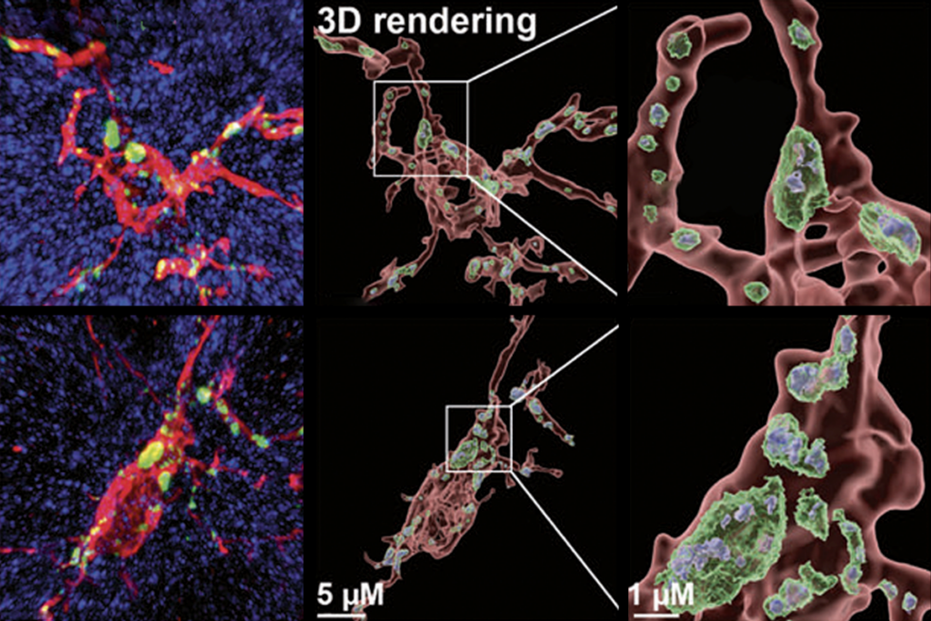
A combination of two microscope techniques enables researchers to record detailed 3-D videos of cells and their structures inside living animals.
A combination of two microscope techniques enables researchers to make detailed 3-D videos of cells and their structures inside live animals1.
The researchers built a custom microscope that combines both methods to produce clear, crisp images. The microscope captures and records images quickly enough to reveal cellular processes in motion.
Each method has limitations on its own, but together, the methods make it possible to visualize details that could previously only be seen in isolated cells or dissected tissue.
One technique, which the researchers developed previously, limits the amount of animal or plant tissue exposed to high-intensity light, which can disturb living cells. Using tunable lasers and filters, the technique, called lattice light-sheet microscopy, shapes light into a thin sheet that quickly passes through the sample, such as a living roundworm or zebrafish. This yields a series of 2-D images that can be combined to make a 3-D video.
But as the light sheet passes through the tissue, the light refracts, or bends. To correct for this, the researchers relied on a technology that astronomers use to remove the atmospheric distortions of distant objects seen through telescopes. They shine a point of light into the tissue they wish to scan, measure distortions in that point and make appropriate adjustments.
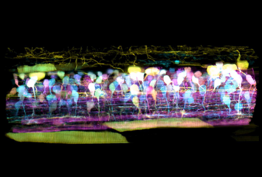
Using their custom microscope, the researchers created short videos within cells and tissues from various organisms and across developmental stages. For example, they were able to observe an immune cell crawl through the inner ear of a zebrafish, engulfing sugar particles along the way. They watched organelles such as mitochondria and the endoplasmic reticulum fragment and reform in dividing cells. And they saw new neurons in the spinal cord of a zebrafish embryo extend long fibers called axons to other cells, forming neuronal circuits.
The researchers also used the method to view cells inside a roundworm, inside leaves from the plant Arabidopsis thaliana and in tissue grown from human stem cells. They described their method 20 April in Science.