This article is more than five years old.
Neuroscience—and science in general—is constantly evolving, so older articles may contain information or theories that have been reevaluated since their original publication date.
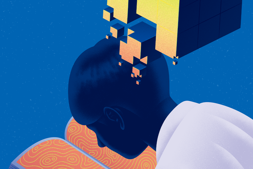
Advanced imaging techniques may reveal more precise pictures of how of the brain’s regions communicate with one another. How much of the neurodevelopmental riddle of autism lies in these tracts?
Today’s news article looks at advances in visualizing white matter, the fibrous tissue of the brain that transmits signals between neurons. Two new imaging techniques stand to reshape our understanding of how the brain’s regions communicate with one another.
Read: New imaging techniques probe brain’s long-range connections »
The new methods are part of the Human Connectome Project, a five-year multi-institutional attempt to map the brain’s wiring. The techniques can render how the lengthy cellular fibers that make up white matter overlap with each other — an important detail that is completely unobservable with previous imaging technologies.
Scientists have long been able to observe neurons and the connections, or synapses, between them, and the focus on synapses dominates studies of neurological dysfunction. However, our window into white matter has thus far been foggy at best.
Long thought to be passive tissue that merely supports neurons, white matter has emerged as a key basis for how the brain learns and functions, by modulating the rate and strength of neuronal signals.
A widely held theory of autism maintains that disrupted long-range signaling is a unifying hallmark of the disorder. A precise picture of how different parts of the brain communicate may prove vital for reliably testing this notion.
What do you think?
Share your thoughts in the comments section below. Or, to dig deeper, continue the conversation in the moderated SFARI Forum for researchers. Not yet a member? Learn how to register here.
Like us on Facebook » | Follow us on Twitter @SFARIcommunity » | Join our newsletter »