The human brain is overwhelmingly complex, and so is the field of neuroscience; insights gleaned from one subfield can be difficult to translate to another. At the same time, interdisciplinary perspectives often spark revelatory new insights into how the brain works and how it can fail. In this series, I speak with neuroscientists who are using interdisciplinary approaches to study the brain, in the hopes of breaking down some of the siloes of neuroscience.
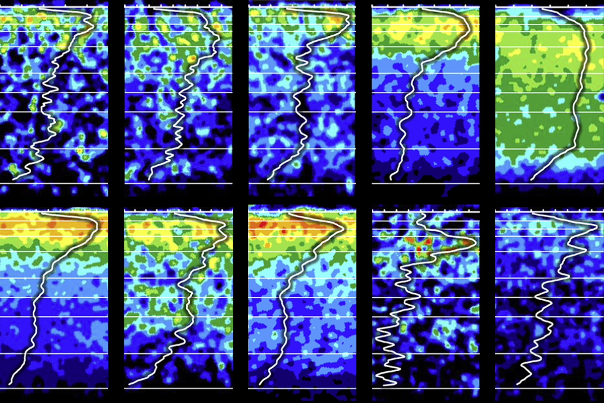
Differing densities: The colored images show the concentration of different receptors in the human primary motor cortex, with high concentration in red and low in blue.
Courtesy of Nicola Palomero-Gallagher and Karl Zilles / NeuroImage (2019)
Add us as a Preferred Source on Google
Set us as a Preferred Source to see The Transmitter more prominently in your Google Search results.
What happens when a histopathologist teams up with computational modelers?
Answers emerge in my chat with Nicola Palomero-Gallagher, a rare example of someone who connects the brain’s microscopic constituents and macroscopic features.
By
Mac Shine
11 December 2023 | 7 min read
Nicola Palomero-Gallagher, a histopathologist at Heinrich Heine University in Düsseldorf, Germany, has an unusual capacity to cross scales. Palomero-Gallagher and her team characterize the human brain in painstaking anatomical detail, describing, for example, the density of neuromodulatory receptors across layers of the cerebral cortex and how that density changes in different regions. These neuromodulatory maps offer an important substrate for exploring how specific neuromodulators shape different cognitive functions.
Few in the field have attempted to connect such microscopic constituents of the brain with its macroscopic network-level expression, making these insights especially valuable. To better understand dopamine’s role in working memory, for example, Palomero-Gallagher teamed up with computational neuroscientists Seán Froudist-Walsh, Henry Kennedy and Xiao-Jing Wang. The interdisciplinary group mapped Palomero-Gallagher’s data on dopamine receptor density onto a finely detailed connectivity model of the macaque cerebral cortex from Wang’s group. The resulting biophysical model recreated activity patterns tied to working memory recorded in macaque brains and revealed how dopamine might help circuits filter out irrelevant information. These findings shed light on how neuromodulators can modulate dynamics across the brain — and demonstrate the benefits of collaborating across fields.
This interview has been edited for length and clarity.
Mac Shine: Deciphering the link between structure and function crosses a few different subfields — neuroanatomy, pharmacology and magnetic resonance imaging. How do you view the role of collaboration in neuroscience?
Nicola Palomero-Gallagher: All my work is highly collaborative, but I’m less interdisciplinary than the young scientists I work with. In order to do good work, I believe that modern students have to go somewhere else, to really work via collaborators to achieve a richer understanding of how to think about brain structure and function.
MS: What inspired you to study neuroanatomy? What questions drive your experiments?
NPG: Well, looking at structure is all well and good, and I’m fascinated by how pretty things can look when you’ve got serially sectioned brains and can detect different structural organizations. But although these pictures are beautiful to look at, there are also limits to the amount of information they provide. To extend our understanding, I try to interpret my results in the framework of functional systems.
I’m a postmortem neuroscientist, meaning that about 99.9 percent of my research has been carried out on histological preparations. Specifically, I look at the distribution of receptors for classical neurotransmitters throughout the human, macaque and rodent brain, using a technique called autoradiography.
Our goal is to bring the receptors into the 3D world by searching for correlations between receptor densities and other views of the brain, such as functional and structural connectivity maps. Or through specific collaborations with computational modelers like Seán and Xiao-Jing.
MS: How can looking at the brain through these different lenses offer new insight into the system?
NPG: Take the example of resting-state functional connectivity [a method to map temporal correlations between brain regions]. We know that the brain is composed of cells and their connections, which we can think of as a semi-rigid scaffold. How might dynamics emerge from this set of relatively rigid structural connections? I’ve been working on the idea that neuromodulatory receptors might play a crucial role here, because they are the ones that enable flexible communication among cells — receptors and neurotransmitters are the bridge that spans that gap between structure and function. Areas that are very far apart in the brain, such as the frontal eye fields and superior parietal lobule, have quite similar receptor expression and can be recruited by the same neurotransmitter.
MS: In neuroscience, we arguably don’t have a great groundwork linking anatomy to physiology. I’m curious as to whether you have strategies that you use with your students and collaborators to help them better understand the anatomy?
NPG: I never liked memorizing lists of names. I always try and associate the structures I study with functions. So, for example, I start with the macroanatomy and link them to global functional frameworks I find compelling.
MS: Another tool people use to make sense of all the anatomical complexity you’re exposing in the brain is computational modeling, but building neural models often requires simplifying the details of the system. There’s a really interesting tension that emerges when you talk to people from computational modeling fields and people studying the brain at the microscopic level, where there is so much exquisite detail. I’m curious to hear what you think of the role of modeling in modern neuroscience.
NPG: From my perspective, anatomists have been around for much longer than modelers, so I think that modelers just haven’t had as long to refine their approaches — I think that’s why modeling just isn’t quite as advanced. Excitingly, I think modelers are starting now to reach the point where they can incorporate neuroanatomy and build more complex models. Like the recent work we’ve done with Froudist-Walsh. In my opinion, it will be the computational neuroscientists who will help integrate all of this information.
MS: How did the collaboration start?
NPG: I met Seán at the OHBM [Organization for Human Brain Mapping] 2017 meeting. He came to see me at my poster after my talk. He introduced himself, said he was a postdoc in Xiao-Jing’s lab, told me a bit about his work, that he had read my work, would love to include receptor data in his models and asked if I would be interested in a collaboration. The story has a surprising element for me because Karl Zilles [a German neuroscientist and anatomist], whom I had worked with on receptor expression studies, happened to be talking to me when Seán came to my poster. It took me some time (actually, weeks!) to realize that it was specifically me, and not Karl, that Seán wanted to meet.
The exchange of ideas at that meeting led to a CRCNS [Collaborative Research in Computational Neuroscience] grant proposal — which we got and has resulted in several articles in high-impact journals! There was quite a budget assigned to travel expenses, as we were supposed to visit each other’s labs at regular intervals. That part unfortunately didn’t happen because of the COVID-19 pandemic. But we had weekly Zoom meetings, and I don’t know if they would have taken place at such regular intervals without the pandemic.
MS: What new collaborations are you excited about?
NPG: I am happy to continue my collaboration with Seán Froudist-Walsh, and to start one with Emmanuel Prozyk, on anatomically informed models of cortical networks. I am also excited about collaborations with Mu-Ming Poo and his team, as well as with Menno Witter, where we aim to better understand structural and functional heterogeneity along the longitudinal axis of the hippocampal formation. I am also planning to expand on cross-species analyses in projects involving Jérôme Sallet, Ting Xu, Rogier Mars and Sarah Heilbronner, where we will combine anatomical and computational expertise to improve our understanding of homologies, which is really important to facilitate translational studies.
Mac Shine
Associate professor of computational systems neurobiology
University of Sydney
See contributor
University of Sydney
Explore more from The Transmitter
Grace Hwang and Joe Monaco discuss the future of NeuroAI
By
Paul Middlebrooks
4 December 2024 | 97 min listen

Imagining the ultimate systems neuroscience paper
By
Mark Humphries
2 December 2024 | 7 min read
How to be a multidisciplinary neuroscientist
By
Austin Coley
15 November 2024 | 5 min read
Cite this article: