Going bad: In many neurological disorders, astrocytes turn from good guys to villains.
Photography by Margeaux Walter

How to teach this paper: ‘Neurotoxic reactive astrocytes are induced by activated microglia,’ by Liddelow et al. (2017)

Shane Liddelow and his collaborators identified the factors that transform astrocytes from their helpful to harmful form. Their work is a great choice if you want to teach students about glial cell types, cell culture, gene expression or protein measurement.

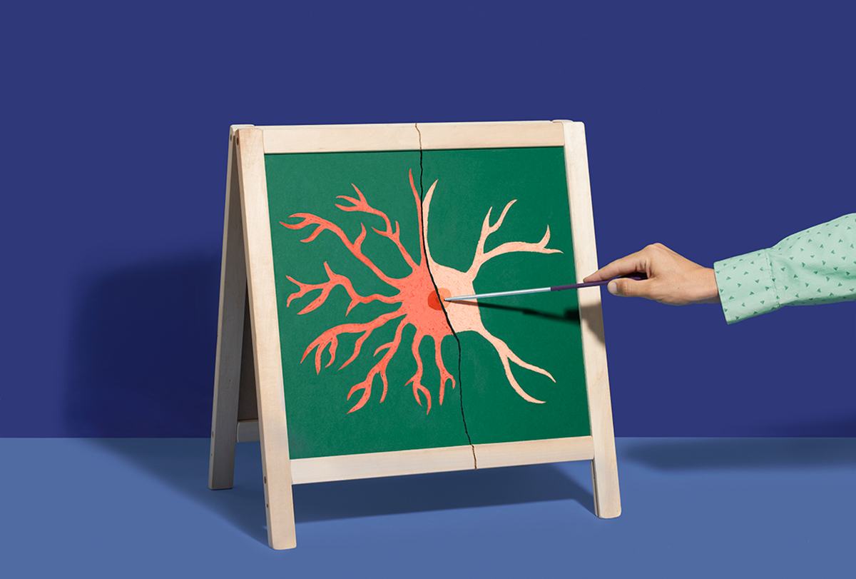
Neurons get most of the hype in neuroscience. But for the past few decades, researchers have been steadily building an understanding of glia, the most numerous cell type in the nervous system. Largely kicked into the spotlight by pioneer Ben Barres and others in the early 2000s, we now understand that glia serve many roles. The friendly neighborhood Spider-Man of the brain, they help to maintain the status quo by secreting growth factors and promoting the formation and function of synapses.

But in response to injury or disease, some glia can transform into cellulae non grata. Specifically, researchers have been interested in how a particular kind of glia, the astrocyte, transforms into a reactive, villainous form, not unlike when Peter Parker goes a bit dark in the movie “Spider-Man 3.”

Although “reactive astrocytes” were first described as early as the 1800s, little was known about how the cells took on this dark form and what happened genetically or functionally when they did. In 2017, a group in Ben Barres’ lab at Stanford University, led by Shane Liddelow, demonstrated that microglia, another form of glia, act as the trigger. Currently cited by more than 5,000 papers, according to Clarivate’s Web of Science, Liddelow et al. (2017) comprehensively showed that these microglia secrete three cytokines that are necessary and sufficient to convert astrocytes from a protective role to a harmful one. The resulting reactive astrocytes then release neurotoxic factor(s) and, in doing so, may play a role in many different neurological diseases. As such, it’s a great paper to elucidate the fine line between helpful and hurtful in the nervous system.

Where this paper might fit

At its core, Liddelow et al. (2017) is a beautiful cellular biology paper. If you want to teach students about glial cell types, cell culture, gene expression or protein measurement, it is a great choice. Although the sequencing techniques in this paper are a bit outdated by 2025 standards—the researchers primarily used quantitative PCR (qPCR)—the use of these simpler techniques gives anyone teaching -omics a chance to talk about more foundational methods.

That said, there are many experiments packed into this paper, which you could take or leave, depending on what your students know (or don’t need to know). For example, the authors used electrophysiology to investigate the function of reactive astrocytes, but you could skip this if it is outside the scope of your course.

Something I love about this paper for an educational context is the immediate link to diseases that students (and the public) will care about. The inclusion of multiple disease states makes this a paper that may even be appropriate for a more clinically focused course, and it opens a path for conversations about how basic neuroscience can be translated into treatments.

Finally, you might use this paper to emphasize the importance of having the right techniques to ask the right questions. Until work that was published in 2011, it was impossible to culture astrocytes in a dish without serum, an artificial way to keep cells alive and one that likely alters astrocytes. The serum-free method, combined with insights from a different lab about the possible transcriptomic differences between astrocyte types, created the perfect foundation for Liddelow to embark on this project.

Working through the figures

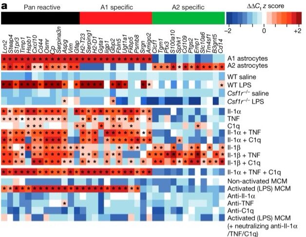
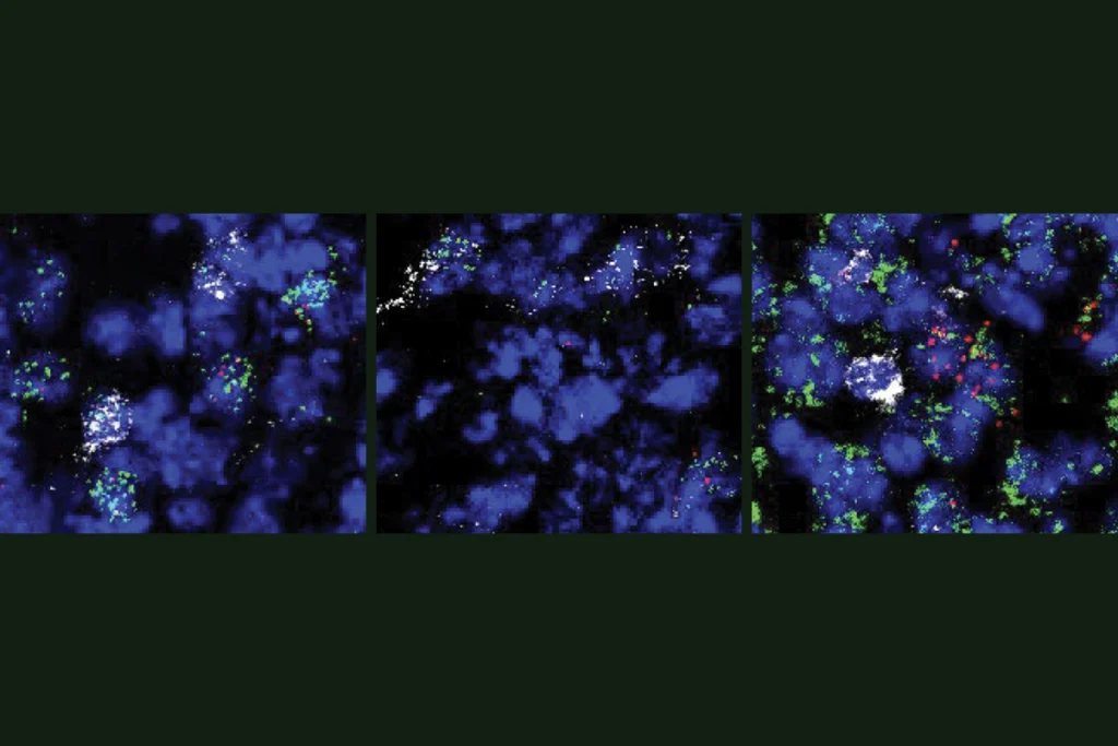
Liddelow and his colleagues began by screening many molecules that they hypothesized would induce reactivity in astrocytes. They then confirmed that a combination of three cytokines was necessary to produce reactivity and that this reactivity didn’t occur in mice without microglia (Csf1r–/-). In a series of experiments that are packed into Figure 1a, they show that both mice and cultured cells without microglia do not produce reactive astrocytes in response to an injection of lipopolysaccharide (LPS). This chemical had previously been shown to induce reactive astrocytes, but astrocytes cannot directly respond to it—they respond only in the presence of microglia.

Finding factors: LPS triggers markers of reactive cells in wildtype (red squares in row 4) but not knockout animals (blue squares in row 6). A combination of three factors, Il-1α, TNF and C1q, can trigger reactive markers, even in nonreactive cells (red squares in row 15.)

Figure 1a itself is a lot to unpack, so let’s break a few features down. First, “A1” (an old name that is no longer used) refers to the neurotoxic reactive substate, and “A2” refers to an alternative state of reactive astrocytes not further studied in this paper. Each row shows a different kind of treatment (often from a knockout animal) that the researchers inspected. Each column shows relative levels of a specific transcript, chosen from the 2011 paper. Red indicates an increase and blue a decrease compared with the control (which changes based on the experiment).

The first major finding—that microglia are necessary to cause astrocytes to take on their harmful form—is illustrated in rows 3 through 6. The second major finding—that the unique combination of three cytokines (interleukin-1 alpha [Il-1alpha], tumor necrosis factor [TNF] and complement component 1q [C1q]) can cause innocent, nonreactive astrocytes to turn reactive in vitro—is seen most clearly in row 15. The authors confirm these qPCR observations using immunohistochemistry and by measuring cytokine secretion directly as well.

The next figure digs into the functional implications of reactive astrocytes by culturing them alongside retinal ganglion cells (RGCs), a type of neuron from the retina that produces many synapses in culture and that the Barres Lab consistently turned to over the years. When RGCs were cultured with reactive astrocytes, the number of healthy synapses decreased, likely because they were producing less of the factors that would normally induce synapse formation. By patching onto RGCs, the authors also demonstrate that the reactive astrocytes caused fewer and weaker excitatory currents. This figure should be fairly straightforward for students who have learned some basics of electrophysiology experiments.

Poor performance: Reactive astrocytes (A1) are worse than controls at trimming synaptosomes (red, top row) and removing myelin debris (red, bottom row).

Returning to their cultured astrocytes, the authors then ask whether reactive astrocytes can perform their typical job of refining neural circuits, by trimming unused synaptic terminals (synaptosomes) or cleaning up myelin debris. Not surprisingly, reactive astrocytes are pretty bad at these things, and they have less of the typical receptors that normally mediate these processes (Figure 3). It’s worth noting that this is the first direct functional study of these astrocytes—although they were often at the scene of the crime, we didn’t know how much of a direct role they were playing.

At this point, the criminal accusations of our reactive astrocytes escalate from petty theft to quite literally murder. When cultured together, reactive astrocytes kill several—but not all— kinds of cells (Figure 4). The good news is that there may be a way to turn this deadly process around. Using a model of optic nerve crush (another oft-used tool in the Barres Lab), the authors show that neutralizing Il-1alpha, TNF and C1q can save the RGCs that would normally die after optic nerve damage (Figure 4). You might challenge students to interpret Figure 4g on their own to transfer what they learned from interpreting Figure 1a. Taken together, the authors conclude that reactive astrocytes are quite literally poisoning the cells around them, releasing some sort of neurotoxin that in the mildest case destroys synapses and decreases electrophysiological activity, and in the worst case, kills them.

A common mark: A gene associated with reactive astrocytes, C3 (shown here in red in both microscopy images and graphs) is upregulated in a number of neurodegenerative diseases.

Optic nerve crush—a great band name, by the way—is not the only scenario in which reactive astrocytes appear. Before this paper, researchers had hypothesized that the transition to reactive may be something that happens in a variety of disease states, as a broad form of neuroinflammation. To test this idea, the authors looked for similar molecular changes in postmortem tissue from people with Alzheimer’s disease, Huntington’s disease, Parkinson’s disease, amyotrophic lateral sclerosis or multiple sclerosis. They found that one of the most uniquely upregulated genes, C3, was indeed present across these samples (and in almost every brain area), giving reactive astrocytes a final, devastating mark on their record: neurodegeneration.

The scientists behind the paper

Lead author Liddelow, now associate professor of neuroscience and physiology, and of ophthalmology, with his own lab at NYU Grossman School of Medicine, recalls the arduous search for a clear transcriptomic signature. “When I would present at lab meeting, I would always start with a slide with a picture of a baby crying, and I would just list things that hadn’t worked for years,” he remembers. In addition to the molecules tested in the main Figure 1, an extended figure lists even more—literally hundreds of molecules in thousands of different combinations.

The story turned a corner after about four years of screening, when Liddelow would repeatedly see Il-1alpha, TNF and C1q emerge. In addition to the extensive screening, the researchers did an “obscene number of control experiments” to confirm their results, says Liddelow, who is proud that their findings have stood the test of time. “Hundreds of studies have validated this in all sorts of different models now, which is really nice,” he says.

Liddelow is still working to better understand reactive astrocytes’ role in health and disease. He has further delineated different kinds of reactive astrocytes, such as those that respond to interferons or those that proliferate. He sounds a bit relieved to have these functionally descriptive names, because the nomenclature surrounding glia, including their use of A1 and A2 in this paper, has long been a bit controversial. The important piece, however, is not the name but the functional characterization of the neurotoxic subtype—something Liddelow looks forward to seeing for more substates of reactive astrocytes discovered by labs around the world.

The 2017 paper ends on quite a cliffhanger—there’s a neurotoxin, but what is it?! Fortunately, the story has continued to evolve. In a 2021 follow-up in Nature, a group in the Barres Lab showed that the culprit is a lipid secreted by astrocytes. These long-chain lipids can be reduced by knocking out the enzyme that synthesizes them, leading to less toxic astrocyte media. Liddelow is working with commercial partners to develop a small-molecule inhibitor of this enzyme. “Twelve years from idea to actual molecule,” says Liddelow, highlighting the often slow pace of translating basic discoveries to clinical relevance. But if this is a core mechanism of brain inflammation, it will be worth the wait.

Additional reading

In addition to Liddelow’s subsequent papers, I would be remiss if I didn’t mention that Liddelow’s postdoctoral adviser, Ben Barres, has a posthumous autobiography that is absolutely worth a read (Barres passed away from pancreatic cancer in 2017). In it he describes the development of the glial research field, details his own growth as a trans scientist, and provides a wealth of advice for budding scientists. The trans and genderqueer students in your class may deeply appreciate the inclusion of these personal details as well—Barres left an important legacy not only for glia, but for the inclusion of all kinds of people in science.

Get alerts for “How to teach this paper” in your inbox.

This series guides educators and self-learners through recent seminal neuroscience papers.