This article is more than five years old.
Neuroscience—and science in general—is constantly evolving, so older articles may contain information or theories that have been reevaluated since their original publication date.

Peruse our picks for the best science photos published on Spectrum this year. We’ve showcased two videos below, too.
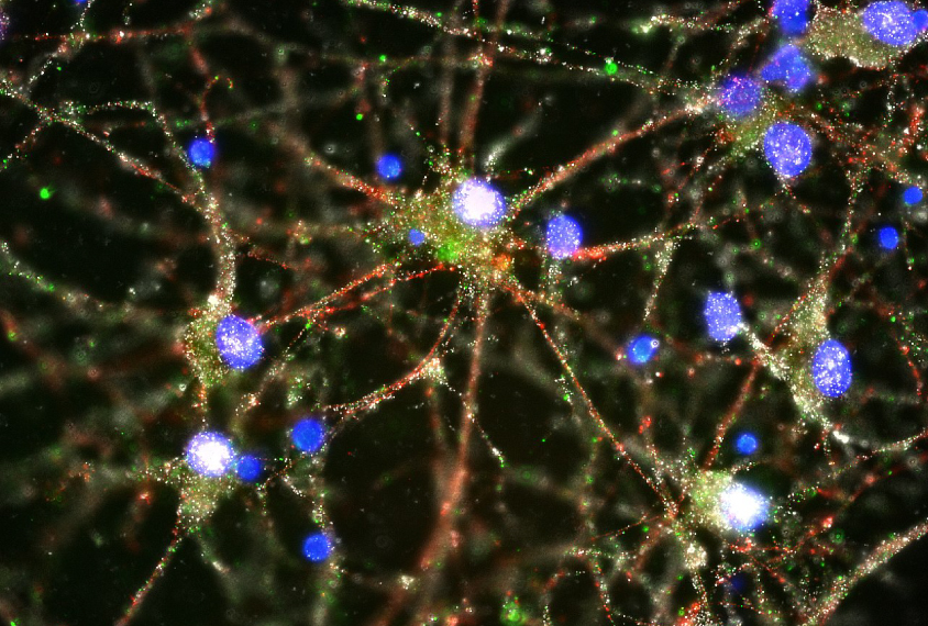
You’re it: ‘Complement’ proteins, in green, tag the junctions of human neurons, targeting them for destruction by immune cells called microglia.
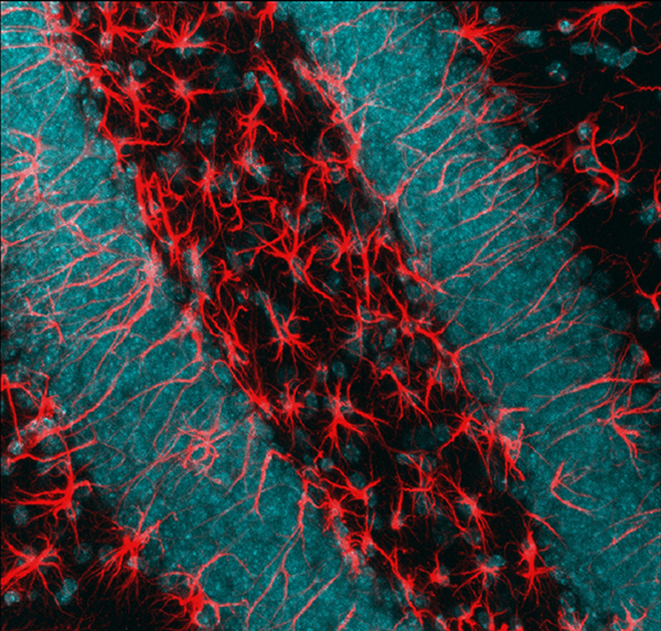
Sorting cells: Cleared brain tissue contains a mix of neurons (nuclei shown in blue) and star-shaped cells called astrocytes (red).
Rainbow connections: Neurons that inhibit brain signals (green) adorn this microscope image of a zebrafish.
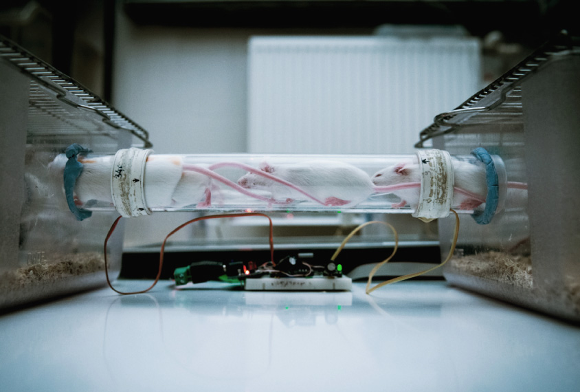
Almost home: A new cage for lab mice mimics the animals’ natural burrows, providing a realistic environment for studies of social behavior.
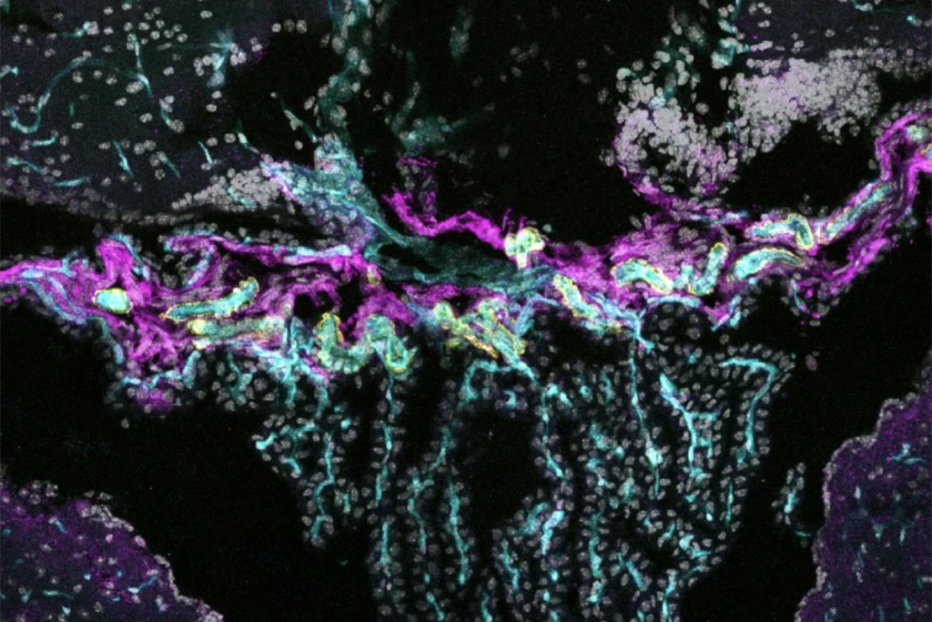
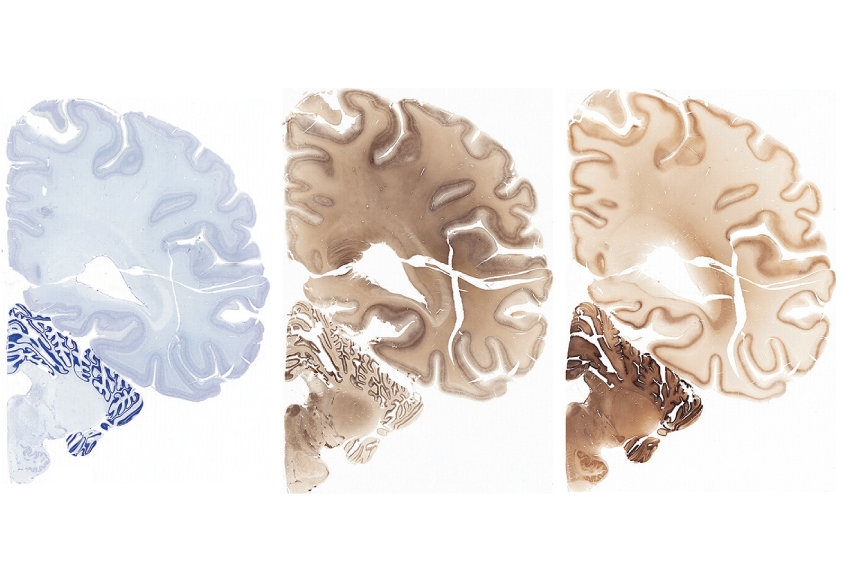
Slices of life: To paint a portrait of a single human brain, scientists colored the tissue with stains that mark certain cells and their parts.
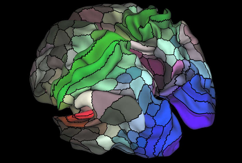
Mosaic mind: A map of the brain’s surface divides each hemisphere into 180 functional areas, including those that govern hearing (red), vision (blue), and sensation and movement (green).
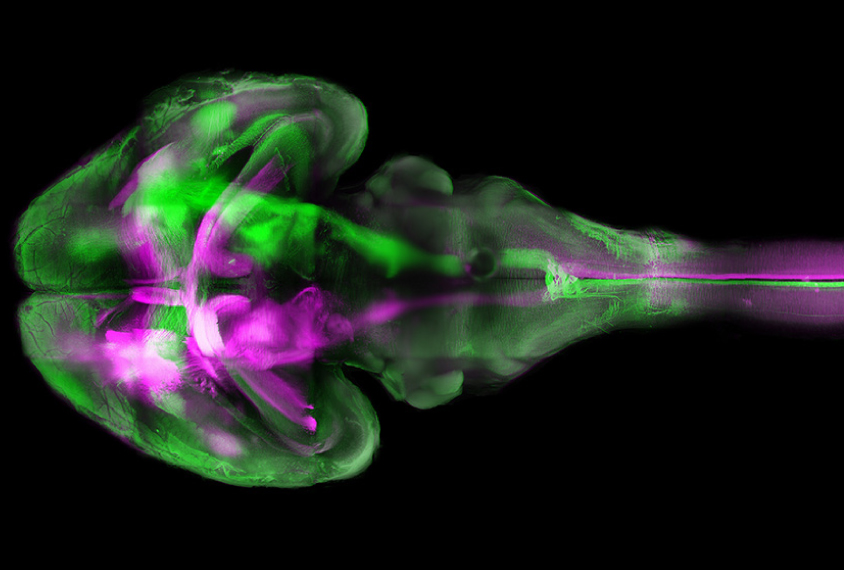
Color cascade: Different nerve tracts glow green or purple in this transparent mouse brain.
Live wires: In this tangle of cells from a tiny piece of mouse brain, a network of neurons (red) spring to action when a mouse sees a certain image.
Baking a brain: Culturing spheres of neurons that resemble the human brain can help scientists understand the effects of the Zika virus or the origins of autism.
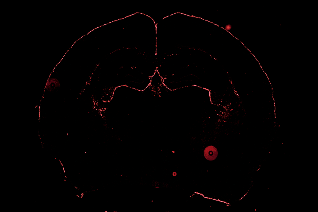
Riot of red: Scientists can turn on a top autism gene, SHANK3 (red), after mouse brains are fully formed.