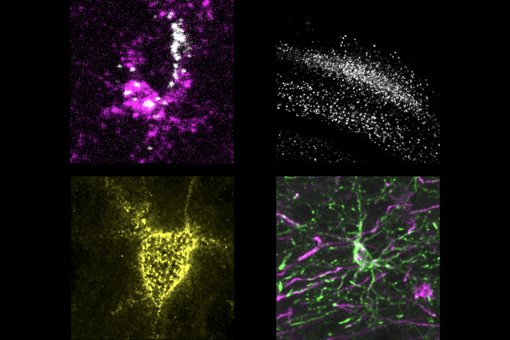
Video: Live from the brain, it’s neuron development
Brain cells communicate across complex junctions called synapses, filled with proteins working to bind neurons together. Kurt Haas of the University of British Columbia in Vancouver has developed a method to watch neuron development in the growing tadpole brain.
Brain cells communicate across complex junctions called synapses. Although synapses are often thought of as the spaces between neurons, they are busy, tightly organized places, filled with proteins working to bind neurons together.
Mutations in several of these proteins have been found in some people with autism, suggesting that errors in the connections that form in the brain could contribute to the disorder.
Because it’s not possible to watch neurons grow in the live brains of typical models like mice, scientists looked for a more compliant animal. Kurt Haas, assistant professor of cellular and physiological sciences at the University of British Columbia in Vancouver, has developed a method to watch neuron development in the growing tadpole brain.
Haas met up with SFARI at the Society for Neuroscience annual meeting in San Diego to discuss the role of two synaptic proteins — neurexin and neuroligin — in autism, and to share his glimpse into a live, growing brain.
For more reports from the 2010 Society for Neuroscience annual meeting, please click here.
Recommended reading
Explore more from The Transmitter

Neuro’s ark: How goats can model neurodegeneration


