This article is more than five years old.
Neuroscience—and science in general—is constantly evolving, so older articles may contain information or theories that have been reevaluated since their original publication date.
A new database contains high-fidelity brain maps of 10 typical adults.
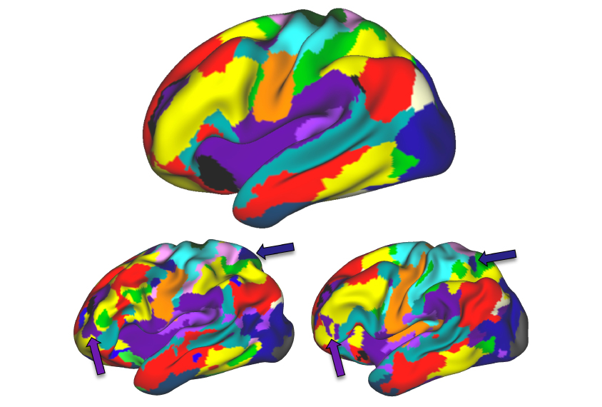
A new database contains high-fidelity brain maps of 10 typical adults1. Each atlas includes neuronal connections particular to an individual rather than averages across the group.
Detailed maps of individual brains could help researchers understand the heterogeneity in autism.
The map of each brain comprises data from five hours of scanning per person, about three times as long as the longest scans in other datasets. The longer the scan, the more reliable the data derived from it2. But the scanners, called functional magnetic resonance imaging (fMRI) machines, are expensive to use, running upward of $600 per hour.
To lower the cost, the researchers took advantage of off-hour pricing — which starts at midnight. They paid $50 per hour, a 90 percent discount.
They scanned each person for six hours while the person made simple movements — of the tongue or feet, for instance — and for at least five hours while the person rested. Participants also underwent 3.5 hours of structural imaging to map their brain anatomy.
Data from the resting-state scans reveal areas of the brain that are spontaneously active. Hours of data can show how brain areas communicate with one another, says lead researcher Evan Gordon, investigator at the U.S. Department of Veterans Affairs.
Brain networks identified at rest correspond with slight differences in the shape, size and location of the regions that become active when individuals make specific movements. This finding shows that variations in brain organization are functionally relevant, Gordon says.
Neurologists may be able to use the technique to diagnose brain conditions. The work appeared 27 July in Neuron.