The red nucleus—a pale pink brainstem structure that coordinates limb movements in quadruped animals—also projects to brain areas that shape reward-motivated and action-based movements in people, according to a new functional imaging study.
The finding suggests the region, like the cerebral cortex, took on a more complex role over the course of evolution. Many researchers had assumed that brainstem structures remained stuck in evolutionarily ancient roles, says Joan Baizer, professor of physiology and biophysics at the University at Buffalo.
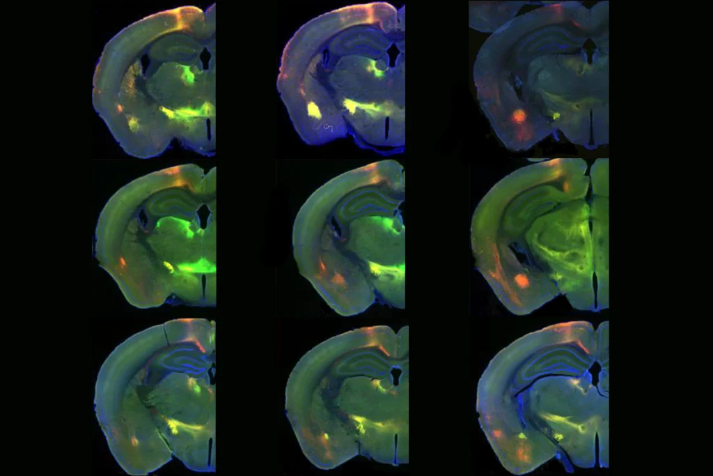
Activity in the red nucleus, a structure that emerged once animals began to use limbs for walking, coordinates the speed and accuracy of those movements in rats and helps to control posture in monkeys, previous electrophysiological recordings have shown. And in nonhuman primates, neurons in the red nucleus project to the motor cortex and spinal cord, anatomical studies have demonstrated, seemingly confirming the area’s role in motor function.
By contrast, the human red nucleus primarily connects to cortical and subcortical regions involved in action control, reward and motivated behavior, the new work reveals.
“If this is such a motor structure, why isn’t it projecting to the spinal cord? That doesn’t really fit with our notion of what this structure is supposed to be doing,” says study investigator Samuel Krimmel, a postdoctoral fellow in Nico Dosenbach’s lab.
The new imaging suggests that, at least in people, the neural underpinnings of motivated movement—previously considered to be the role of higher-order brain areas—reach “all the way down into the brainstem,” says Dosenbach, professor of neurology at Washington University School of Medicine, who led the work. The findings were published last month in Nature Communications.
T
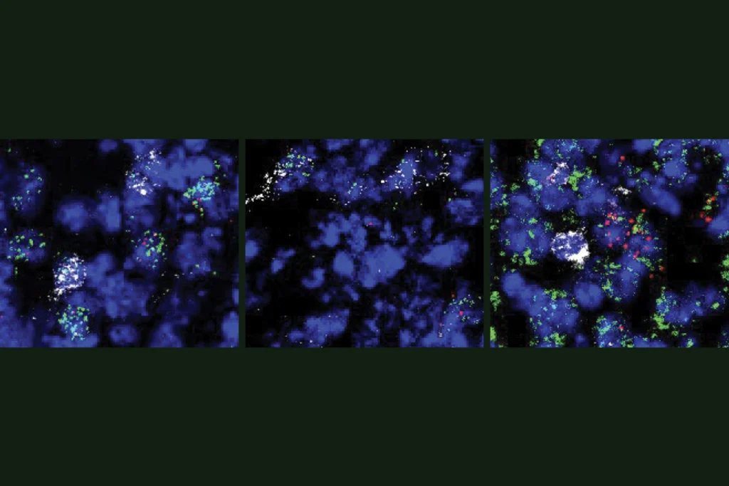
he red nucleus is made up of two subregions: the magnocellular red nucleus, which projects directly to the spinal cord; and the parvocellular region, which connects to other parts of the brain, including the cortex. In quadruped animals, such as reptiles and felines, most red nucleus neurons are magnocellular, which may explain why the region has long been characterized as a motor structure, Krimmel says.As animals evolved from quadrupedal to bipedal, the ratio of red nucleus neurons changed. At least half of the neurons in the red nucleus of nonhuman primates, for example, are parvocellular, some work suggests. And in humans, most are parvocellular neurons. But anatomy aside, pinning down the structure’s function in people has proved historically difficult; the brainstem has a low signal-to-noise ratio that makes it challenging to image with functional MRI (fMRI), Krimmel says.
To work around that challenge, the team analyzed repeated brain scans they had previously collected from five people. With this approach, called precision functional mapping, researchers collect large amounts of fMRI data for a few participants rather than relying on a small amount of data from many people.
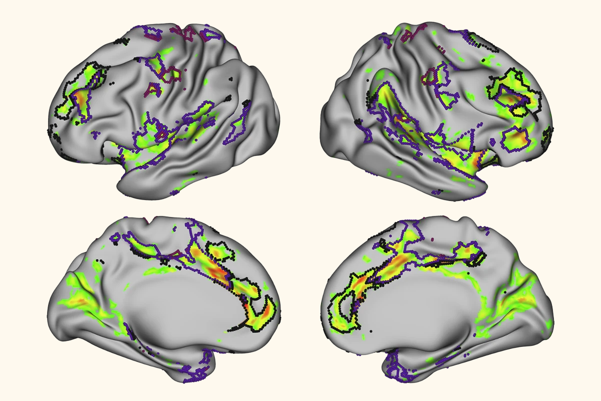
Many of the regions targeted by projections from the human red nucleus belong to the salience and action-mode networks, which respectively coordinate motivated behaviors and actions, the analysis showed. For example, the red nucleus projects to the dorsal anterior cingulate cortex, shown to be involved with reward processing. Those findings held up in group-averaged data from the UK Biobank, the Adolescent Brain Cognitive Development Study and the Human Connectome Project.
Although the red nucleus does connect with the motor cortex, those signals are limited to areas of the cortex that are a part of the somato-cognitive action network (SCAN), a network that Dosenbach and his colleagues identified in 2023 and that coordinates goal-directed body movements.
D
espite the limited number of participants, the study’s use of repeated scans helps to validate the findings, says Steve Nelson, associate professor of clinical behavioral neuroscience at the University of Minnesota Twin Cities, who was not involved in the work. “You can do an experiment in an individual, and then you can see if that replicates in another individual.”Still, the work cannot conclusively answer questions of how the red nucleus shapes behavior, Nelson says. Future studies that include reward-based tasks, for example, could help better characterize the region’s functional role, he adds.
That would also help narrow down which behavioral aspects of “reward” and “motivation” the red nucleus is involved in, says Cedric Boeckx, research professor in the evolution of human cognition and language at the Catalan Institution for Research and Advanced Studies, who was not involved in the work.
“We could look at the structure to see exactly what it does and how it essentially interacts with other brain regions to perform that role,” Boeckx says.
Researchers should also investigate the function of other brain areas thought to be conserved across evolution, but which may have changed over time, Boeckx adds.
That idea is already on Krimmel’s radar. “We should focus on the structures where we know what they do—quote unquote, ‘know’—because I think those are areas where we’re probably wrong,” he says.