Listen to this story:
0:00
/
Human neuroscience research has largely overlooked this spatial scale—which bridges cells and brain areas. But new advances in functional MRI technology are changing that.
The brain is a network organ, comprising an immense number of cells that send neuronal signals across connected subsystems. Traditional tools for studying those systems focus on the microscopic scale, measuring electrophysiological signals from individual brain cells, and the macroscopic scale (centimeters)—capturing activity across the whole brain with functional magnetic resonance imaging (fMRI), typically resolving 100 to 200 brain areas at once. But the mesoscopic, or “middle,” scale that bridges these two extremes has been largely overlooked. Electrophysiology, though precise, is invasive and difficult to apply on a broad scale, and traditional MRI is often too noisy to resolve smaller brain structures. As a result, traditional methods fail to provide the spatial resolution needed to connect microscopic processes to macroscopic brain networks.
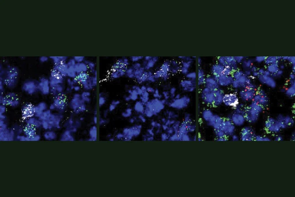
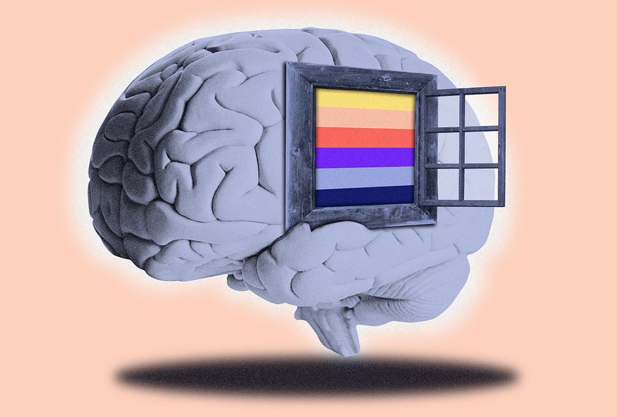
This mesoscopic scale, however, offers a fascinating window into the brain’s circuitry and connectivity. At this level, we can resolve the basic building blocks of neural information processing: the cortical layers, which house different types of neurons. Invasive animal studies and microscopy of cadaver samples show that each layer performs specialized computations and connects to other brain areas in distinct ways. In the frontal lobe, for example, the upper cortical layers integrate input from other cortical areas, and the deeper layers handle output, such as triggering muscle movements. In sensory systems, such as the visual, auditory and somatosensory cortices, the middle layers process input from the senses. Meanwhile, the upper and lower layers handle internal connections from higher-level brain regions based on expectations, attention and stimulus context. By pushing the resolution of fMRI from conventional centimeter-wide blobs of activity down to the level of cortical layers, we can do much more than just see whether a brain area is active. We can figure out how it is involved, where activity is coming from and why it is being modulated.
Technological advances over the past 10 years are making it possible to explore the brain at this vital level—an approach known as layer fMRI. MRI hardware with field strengths of 7 tesla; new signal readout techniques, including accelerated multi-shot echo planar imaging; and newly established MRI contrast methods, such as vascular space occupancy, or VASO, have enabled researchers to push resolutions higher than ever before. Today, most modern fMRI scanners can capture neural activity changes down to the mesoscopic scale—the level of layers—granting unprecedented access to the network organization of the brain.
This type of research will be especially important for studying developmental and psychiatric conditions, many of which have a network component. To understand their causes and to develop treatment tools, we need to look at them from the perspective of network disruptions, at a spatial scale that resolves changes in information flow within the network—namely, the mesoscopic scale.
T
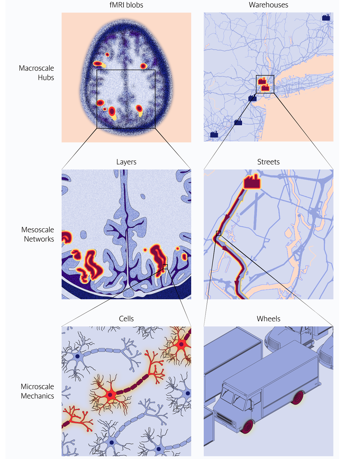
o illustrate the importance of this middle level, let’s use a human network everyone knows: freight transportation. In this network, a traffic jam can block highway lanes, disrupting connectivity between hubs and delaying goods from arriving on time. Now imagine we’re researchers investigating this disruption across different spatial scales. At the macroscopic scale, we would see less activity at the big hubs—major warehouses. This reveals a problem with the network, but not the cause. At the microscopic scale, we might zoom in and see trucks’ stationary wheels. This captures the mechanical underpinnings of the network disruption, but it doesn’t reveal the root reason—traffic congestion. To actually fix the problem, we need information from the mesoscopic transportation scale. At that level, we can identify which routes are congested and how this is disrupting traffic flow, enabling us to propose traffic rerouting to alleviate the issue.This same logic applies to networks in the brain. At the macroscopic scale, conventional fMRI lets us observe activity fluctuations between brain areas, giving insights into functional connectivity. However, it doesn’t tell us why connectivity is disrupted. At the microscopic scale, techniques such as electrophysiology dive into the underlying mechanisms of these disruptions and detect the changes in activity in individual cells. But they lack the broader coverage to place this activity in the context of the whole network. Layer fMRI bridges this gap by revealing specific cortical layers involved and the directionality of connectivity issues.
Use of layer fMRI has grown rapidly since 2016. Ultra-high-field 7 tesla scanners, which are more than twice as strong as most hospital scanners, are particularly well suited for layer fMRI. And they are growing in popularity, with more than 125 scanners installed worldwide. However, challenges remain: Most 7 tesla MRI scanners were designed for high-resolution clinical imaging rather than layer fMRI. That means that layer fMRI still requires technical expertise and adjustments in retrofitting scanning procedures, challenging the technique’s scalability.
Fortunately, researchers are addressing these issues, developing standardized analysis pipelines that are user-friendly for non-experts and adapting acquisition protocols for lower field strengths, such as 3 tesla. These ongoing advances have the potential to increase the widespread use of layer fMRI across various disciplines and imaging centers. Improvements in imaging technology, such as stronger MRI magnets and better equipment, including gradients and RF coils, will soon allow us to see the cortical layers in even further detail. These advances won’t just enable us to study the brain more precisely, they will also help scientists better understand the neurovascular underpinnings of fMRI signals.
I hope that the field of layer fMRI fulfills its potential to bridge the gap between preclinical invasive animal brain research and human cognitive neuroscience. Only with further cross-disciplinary research efforts that cover the right spatial scales will we be able to understand the brain as the network organ it is.