Neurons in the visual cortex decode an object’s orientation—horizontal, vertical or anything in between—using information from non-orientation-tuned neurons in the thalamus, according to David Hubel and Torsten Wiesel’s Nobel Prize-winning work in cats in the 1950s and ’60s. In other species, though, the process remained unclear. Thalamic neurons in mice, for example, show orientation selectivity, subsequent studies suggested.
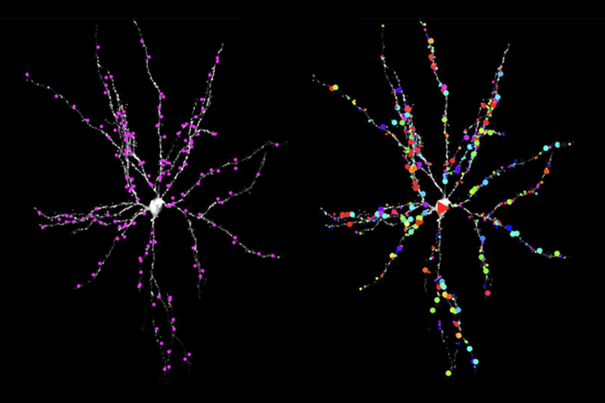
New mouse findings—realized by imaging individual synapses on cortical neurons and distinguishing which inputs come from the thalamus versus the neighboring cortex during visual processing—help resolve the discrepancy. Signals coming into the primary visual cortex, or V1, from the thalamus are not orientation tuned, but those from other parts of the cortex are, confirming that orientation tuning occurs in the visual cortex, the new study reveals.
This study is the first “to get a map of thalamic receptive field location at the level of seeing almost all the spines that receive thalamic input,” says Jose Manuel Alonso, professor of biological and vision sciences at the State University of New York College of Optometry, who was not involved with the work. “This is unbelievably beautiful.”
What’s more, the Hubel and Wiesel model of orientation selectivity “is preserved through evolution,” Alonso adds.
“In the mouse, this pathway from the thalamus to the V1 is really organized as the Hubel and Wiesel suggested it should be,” says Anton Arkhipov, investigator at the Allen Institute, who was not involved with the study.
Thalamic neurons responded broadly to all orientations of alternating black and white lines in a video, glutamate imaging and optogenetic manipulation in the new study demonstrate.
Importantly, the thalamocortical connections showed no significant responses via calcium imaging, which could have implications for both past and future studies, Alonso says. “If you are interested in measuring thalamic inputs to the cortex in vision in the mouse, you cannot use calcium imaging. You have to use glutamate imaging.” Additionally, “all the assumptions that were made based just on calcium imaging have to be reexamined.”
T
he study investigators initially tried to use calcium imaging to monitor orientation-tuned neurons in the visual cortex, but they found something surprising, says Marinus Kloos, who conducted the work as a Ph.D. student at the Technical University of Munich in the lab of Arthur Konnerth. Even though these neurons had spines that received signals from both the neighboring cortex and the thalamus, according to tracing experiments, only the former turned on during visual stimulation, whereas the latter remained silent.“We did not see these typical thalamic inputs with calcium imaging,” says Kloos, who is now a postdoctoral researcher at Ludwig Maximilian University of Munich.
Activity at thalamocortical synapses appeared only when Kloos and his colleagues used glutamate imaging. And the synapses in the visual cortex that have an orientation preference involved neurons elsewhere in the cortex, not in the thalamus, the researchers found using optogenetics.
This orientation selectivity also influenced the synapse location, as cortical synapses with similar orientation preferences tended to cluster in the dendrites of neurons in the visual cortex. On the other hand, thalamic inputs responded to all stimulus orientations without any preference, and they distributed evenly throughout the dendrites.
According to Hubel and Wiesel’s model, orientation tuning stems from the combination of inputs from thalamic cells that activate when light hits the center of their receptive fields (ON-center fields) and inputs that respond to darkness in their field’s center (OFF-center fields).
When Kloos mapped the receptive fields of all thalamic inputs onto one V1 neuron, the ON-center and OFF-center fields arranged in an ellipsis that matched the neuron’s preferred orientation. This pattern also held true for five other neurons. “If you have a bar moving in exactly that orientation, you will have perfect activation of all the thalamocortical synapses at the same time, and this can really then drive the cell to fire,” Kloos says. “It perfectly matches up with what Hubel and Wiesel hypothesized.”
T
he findings, published in March in Science, put to rest any concerns about using mice in the field of vision, Kloos says. “The visual system of the mouse is really suitable to study the visual system in general, because there are very basic mechanisms that are still true.”But there are still many unknowns, he adds. Even in layer 4 of the V1, the focus of the new study, there are many more cell types, such as different subclasses of inhibitory neurons, Kloos says. Ultimately, the field needs a full picture of all the cell types that connect the thalamus and V1, he adds.
“What they found is very important. It’s foundational,” Arkhipov says. “It proves what many people thought to be happening, but it still does not provide, really, a complete picture.”
Questions also remain about why the activation of thalamocortical synapses does not produce postsynaptic calcium events and what that might mean for learning, Kloos says. For example, it could be that, instead of activating NMDA receptors, visual stimulation activates only AMPA receptors, which are not permeable to calcium, he speculates. “It could mean that those are synapses [that] are just very robust, and they don’t need to go [through] any plasticity.”
For vision researchers, “it also means that all the synapse imaging done with calcium imaging is perfectly fine, perfectly correct,” Kloos says. “It’s just the possibility that you [might] miss something.”