When artificial neural networks learn new information, their older memories sometimes vanish. This process, known as catastrophic forgetting, plagues computational models of the brain. How the human brain avoids it is a long-standing puzzle.
A new modeling study begins to fit the pieces together: It proposes a realistic way for multiple, overlapping memories to form and coexist, though this mechanism still needs to be confirmed experimentally.
“We have somas, dendrites, neurons that are inhibitory and excitatory,” which makes the model “biologically plausible in the way that [artificial neural networks] are not,” says study investigator Julijana Gjorgjieva, professor of computational neuroscience at the Technical University of Munich.
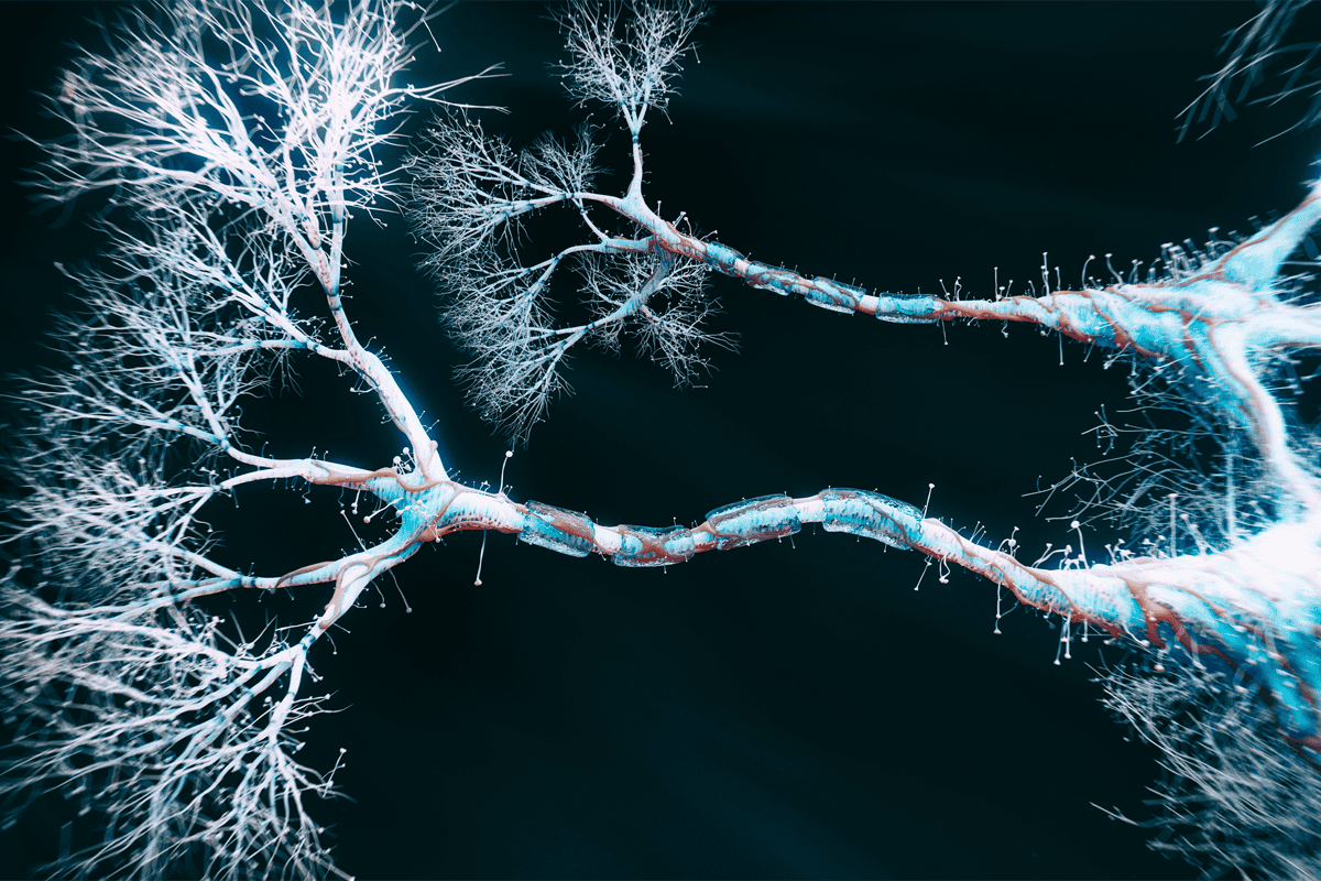
Dendrites act as separate, semi-independent compartments within a single neuron, the study suggests. Inhibitory interneurons selectively unlock learning in one compartment at a time while keeping the others unchanged. This allows the same neuron to join multiple assemblies—groups of neurons that represent thoughts or memories—without erasing what it learned previously. The study was published in Neuron in August.
This work is “a major advance” toward figuring out how the spiking of individual neurons gives rise to complex behavior, says Christos Papadimitriou, professor of computer science at Columbia University, who was not involved in the work. Assemblies, not single neurons, are increasingly seen as the brain’s basic computational units, Papadimitriou says.
He previously theorized that assemblies could, in principle, carry out any computation by copying themselves or merging with other assemblies. This theory was “biologically inspired; this paper renders it biologically realized,” he says. “I was very impressed with the thoroughness and how imaginative [the study] is. It’s something that I was hoping somebody would do.”
T
he researchers simulated a network in which each neuron connected to every other neuron. Each neuron had a soma and multiple dendritic compartments that could each independently generate NMDA-receptor-driven spikes and strengthen their connection to other neurons. Inhibitory neurons selectively silenced individual dendritic compartments, blocking plasticity from occurring there but leaving the others untouched.The researchers applied one of several context signals, each of which silenced a subset of the inhibitory neurons in the model, to mimic what happens when an animal enters a new environment or internal state. These signals reduced the inhibition on select compartments, allowing learning to happen.
Then, various groups of excitatory neurons activated the network, mimicking what happens when an animal views a series of images. Each group generated a different assembly, akin to forming a memory of that image; activating the same group in a different context produced separate assemblies.
After an assembly was formed, if only a subset of these excitatory neurons activated the network—like when an animal sees only part of an image—the full assembly joined in, but only if the reactivation happened in the same context in which that learning occurred.
The researchers modeled multiple brain areas as recurrently connected networks linked to one another only by feedforward connections. The model could send a copy of an assembly from one area to another, the study showed. If two different areas both projected to a shared downstream area under the same context, the downstream area combined the patterns from the two upstream areas into a single joint representation.
This process represents something like combining information from multiple modalities, such as binding an image and sound together into a single concept, says study investigator Christoph Miehl, a postdoctoral fellow in Tim Vogels’ lab at the Institute of Science and Technology Austria. These associations formed in two distinct ways, the team found: simultaneously, in which both areas activated at once and formed a new assembly downstream; or sequentially, in which a new association attached to an already-existing assembly.
Single neurons “are really important for computations on the big level, like combination of information across areas. That’s the beauty of our work: We bridge across these two different scales,” Miehl says.
In a simulated task, the model successfully distinguished visually similar but phonetically distinct characters, such as “0” and “O.” Visually similar pairs of stimuli activated largely overlapping assemblies in a simulated visual area of the brain. But in a downstream “concept” area that received both visual and auditory input, similar stimuli ended up in nonoverlapping assemblies. This allowed the concept area to correctly separate stimuli that the visual area alone could not, even if the concept area just saw the image without sound, as it received the right context.
T
he model contains several simplifying assumptions, says study investigator Sebastian Onasch, a former graduate student in Gjorgjieva’s lab. It assumes, for example, that the inhibitory population connects to only one dendrite of each neuron in a specific context, whereas real inhibitory connectivity is likely more complicated. This assumption does not need to hold for the model to be informative, but “without this context-dependent inhibition, you lose a little bit of this ability to segregate stimuli,” Gjorgjieva says.But the model does offer testable predictions about interneuron organization, says Attila Losonczy, professor of neuroscience at the University of Texas Southwestern Medical Center, who was not involved in the work. Distinct subgroups of an inhibitory cell class should respond to distinct contextual signals, those subgroups should preferentially target specific dendritic branches, and dendritic activation patterns should shift systematically with context.
And Losonczy’s recent findings support the idea that different parts of the dendrite make different computations. Distal and proximal dendrites can decouple from each other, with distal dendrites encoding contextual information.
Dendrites in hippocampal and cortical neurons have tiny constrictions that partition them into distinct electrical compartments, according to a paper published in Science Advances last month. These constrictions could provide the physical mechanism by which dendrites compartmentalize, Gjorgjieva says. The constrictions alone can’t explain the context-dependent assembly formation, however—that requires the inhibition and disinhibition of specific compartments. But it provides “anatomical support for the broader idea that dendrites contain local computational and plasticity compartments,” she adds.
The biggest open question is what “context” actually is in the brain and where it comes from, Miehl says. In the future, the researchers say they would like to move beyond the model’s simple visual-auditory task and closer to a reward-based behavioral task that a mouse might solve.
The study “provides us with a novel function of dendritic inhibition,” Losonczy says. “It provides a really strong testable hypothesis of what comes next.”