Research image of neuronal migration.
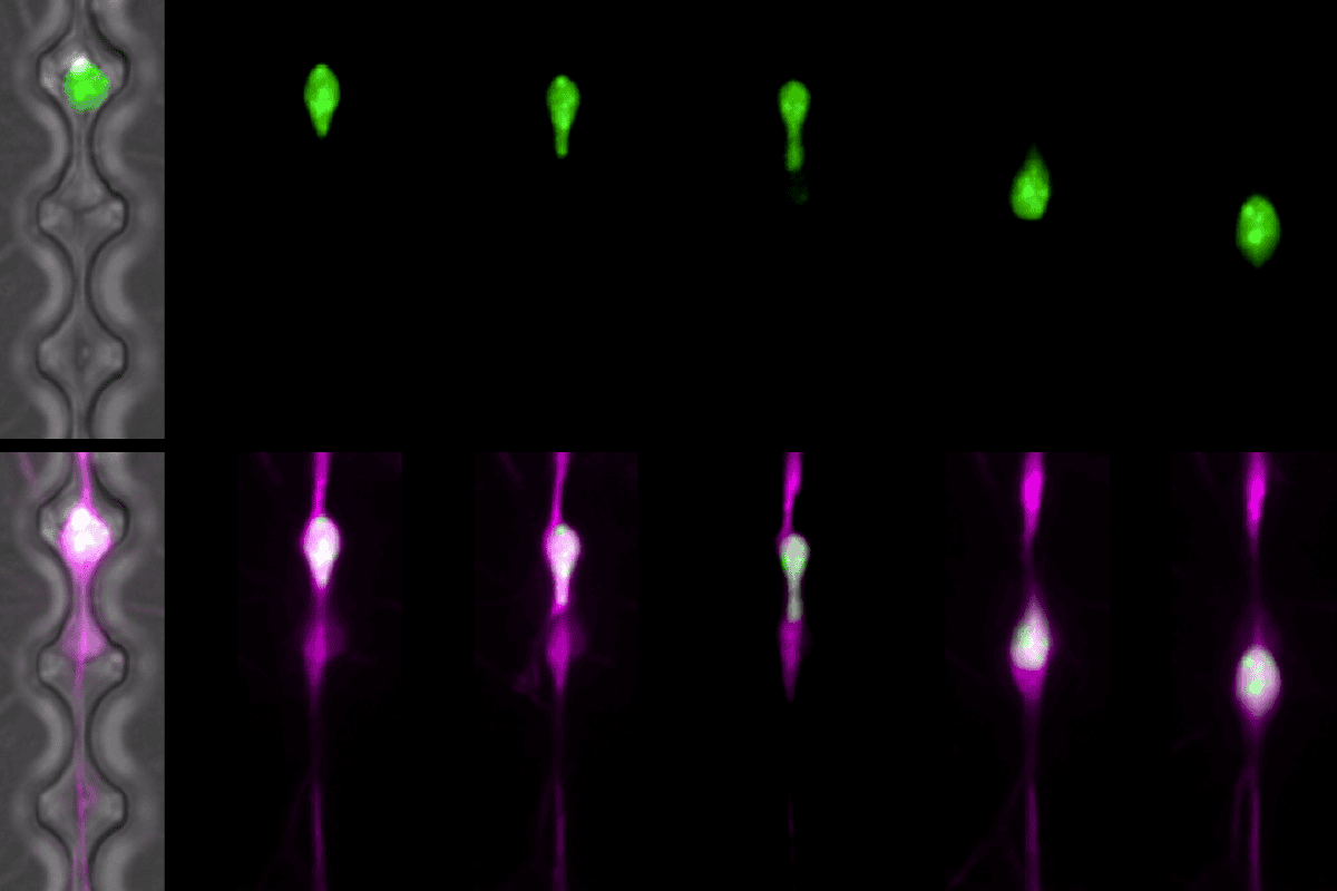
Tight squeeze: The nucleus (green) of cerebellar granule neurons (pink) gets squashed as the cells flow down chips with 3-micrometer-wide channels, imitating neuronal migration.
Photography by the Kengaku Lab
Add us as a Preferred Source on Google

Neurons engage in risky, DNA-breaking business during migration

The cells later repair the damage, which occurs mostly in transcriptionally silent parts of the genome.

By Sarah Thau
24 July 2026 | 4 min read

Neuronal DNA breaks as the cells migrate during development, though enzymes later fix that damage to preserve function, according to a study published in Nature last month. 

“DNA damage is something that you think about when you might have an X-ray or when you go flying in a plane,” says David Rowitch, deputy director of basic and translational research at Cedars-Sinai Guerin Children’s and professor of pediatrics at the University of Cambridge, who was not involved in the study. “What people maybe don’t understand as well is [that] during development, there’s a lot of DNA damage that normally occurs.” 

During human fetal development, neurons travel from the ventricular zone to take up residence elsewhere in the brain and nervous system. This trek “is a risky process; neurons seem to have a very dangerous trip,” says study investigator Mineko Kengaku, professor of developmental biology at Kyoto University’s Institute for Integrated Cell-Material Sciences.  

The nuclei of migrating cells can become deformed as the cells pass through tight intercellular spaces, previous work shows. In cancer and immune cells, this migration can rupture the nuclear envelope. That doesn’t happen in neuronal nuclei, but they still elongate, rotate and distort during their travels, according to the live-cell imaging presented in the new work.

Kengaku and her colleagues forced fetal mouse cerebellar granule neurons to squeeze through microfluidic channels etched into a 2D chip, approximating the cells’ journey in vivo. Fluorescent tags marked the cells’ nuclear envelope and DNA damage in distinct colors. 

As the nucleus of each cell is distorted during the cells’ passage, most acquire double-stranded DNA breaks, the study shows. An enzyme called topoisomerase 2-beta covalently binds the breaks, which the cells then mend via non-homologous end-joining using the enzyme ligase 4, the primary mechanism by which cells repair breaks without a template strand. 

Cerebellar granule neurons from mice missing the repairing ligase changed their expression of only 336 genes. These knockout mice developed normally, aside from subtle differences in their motor functions later in life. The results suggest that “DNA damage is inevitable,” Kengaku says, but the neurons have a “mechanism to make the DNA damage in safe genomic regions.”
Time-lapse traveling: Cerebellar granule neurons (pink) squeeze through microfluidic channels etched into 2D chip, but their nuclear envelopes (green) do not rupture.
Video by the Kengaku Lab

S

till, the behavioral deficits are “a signpost to a permanent physiological problem,” Rowitch says. “We don’t quite appreciate the extent to which failure of DNA repair plays a role in manifestations of human neurodevelopmental disease. When it comes to neurodegenerative disorders, this is probably even less well understood.” The importance of DNA repair mechanisms is “a message which I think we’re going to start to see in a variety of neurodevelopmental syndromes,” he adds.  

The study used cerebellar granule neurons because they are the most abundant neuron type in the brain, and “they are most easy to handle” compared with cortical excitatory neurons and cortical interneurons, which are the other two models used to study migration, Kengaku says. But “it’s unclear if they’re representative of other parts of the brain and how they develop,” Rowitch says, leaving room for further study. 

What’s more, the work “prompts us to continue to work on the tools to be able to see the genome in the cellular context,” says Mercedes Paredes, professor in residence of neurobiology and neurology at the University of California, San Francisco, who was not involved in the study. 

By using a wide range of tools and new technologies to look at migrating neurons, “we can learn a lot about how neurons evolve such fantastic functions, by taking a big risk,” Kengaku says. She next wants to dig deeper into the biomechanics of the nuclear envelope and “what makes the difference between other types of cells, like cancer cells and immune cells, which undergo rupture?”

Sign up for our weekly newsletter.

Catch up on what you missed from our recent coverage, and get breaking news alerts.