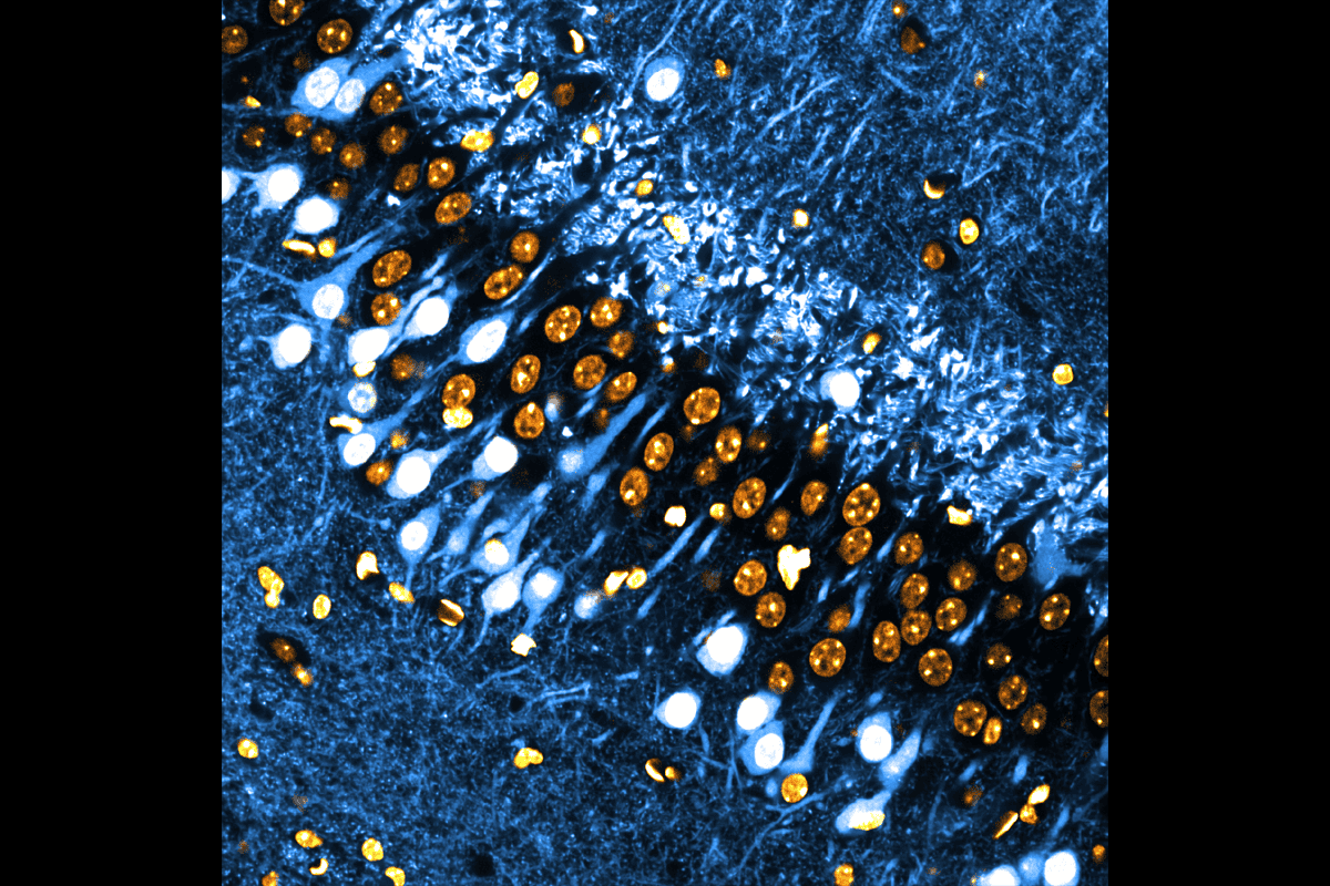
Two of a kind: The CA3 region of the mouse hippocampus has two types of pyramidal neurons, according to a new preprint: one type (shown in blue and white) expresses the transcription factor ST18, while the other one (with only the nuclei visible here, in orange), does not.
Add us as a Preferred Source on Google

Textbook model of hippocampus’s role in memory may need updating

The CA3 region, long thought to have just one circuit, has two, sporting different wiring and functions, a new preprint suggests.

By Alissa de Chassey
4 August 2026 | 5 min read

A long-standing model of the hippocampus’s role in memory needs to be revised, according to a new preprint

For more than half a century, memory theories treated the CA3 region of the hippocampus as a uniform population of pyramidal neurons that form one broad recurrent, or autoassociative, network; the cells synapse onto each other and also send signals to the CA1 region. The network stores memories as synapses strengthen among coactivated cells, each encoding a different piece of the memory. And because of this architecture, a partial cue can reactivate a full memory, such as when the taste of a madeleine sparks a flood of childhood memories for the narrator of Marcel Proust’s “In Search of Lost Time.”

But it turns out that picture may be wrong. The CA3 instead comprises two distinct types of pyramidal neurons arranged in two layers, with different morphology, physiology and connectivity patterns, the preprint suggests. The findings were posted on bioRxiv in July.

“These two cell types are very different, and one of them is totally breaking what the textbook would say,” says study investigator Jake Watson, a postdoctoral researcher in Peter Jonas’ lab at the Institute of Science and Technology Austria. 

A single transcription factor, ST18, distinguishes the two populations, the study reveals: A set of superficial CA3 neurons that express ST18 forms a recurrent network as predicted by the classical model, and a deeper set, which does not express ST18, regulates the superficial one.

This configuration “splits the information into two computational processes, and two sets of neurons are doing something different,” says Gyorgy Buzsaki, professor of neuroscience at New York University, who was not involved in the work.

Having identified genetic markers of a potential separate cell population, whether the CA3 or a more general deep hippocampal layer, will transform how we think today about this brain region critical for memory,” says Liset M. de la Prida, professor and leader of the Neural Circuits Laboratory at the Instituto Cajal, who was not involved in the study. “I think it will transform how [the CA3] processes information and receive the inputs and integrate the inputs, it looks like [the two populations] could be playing different roles.”
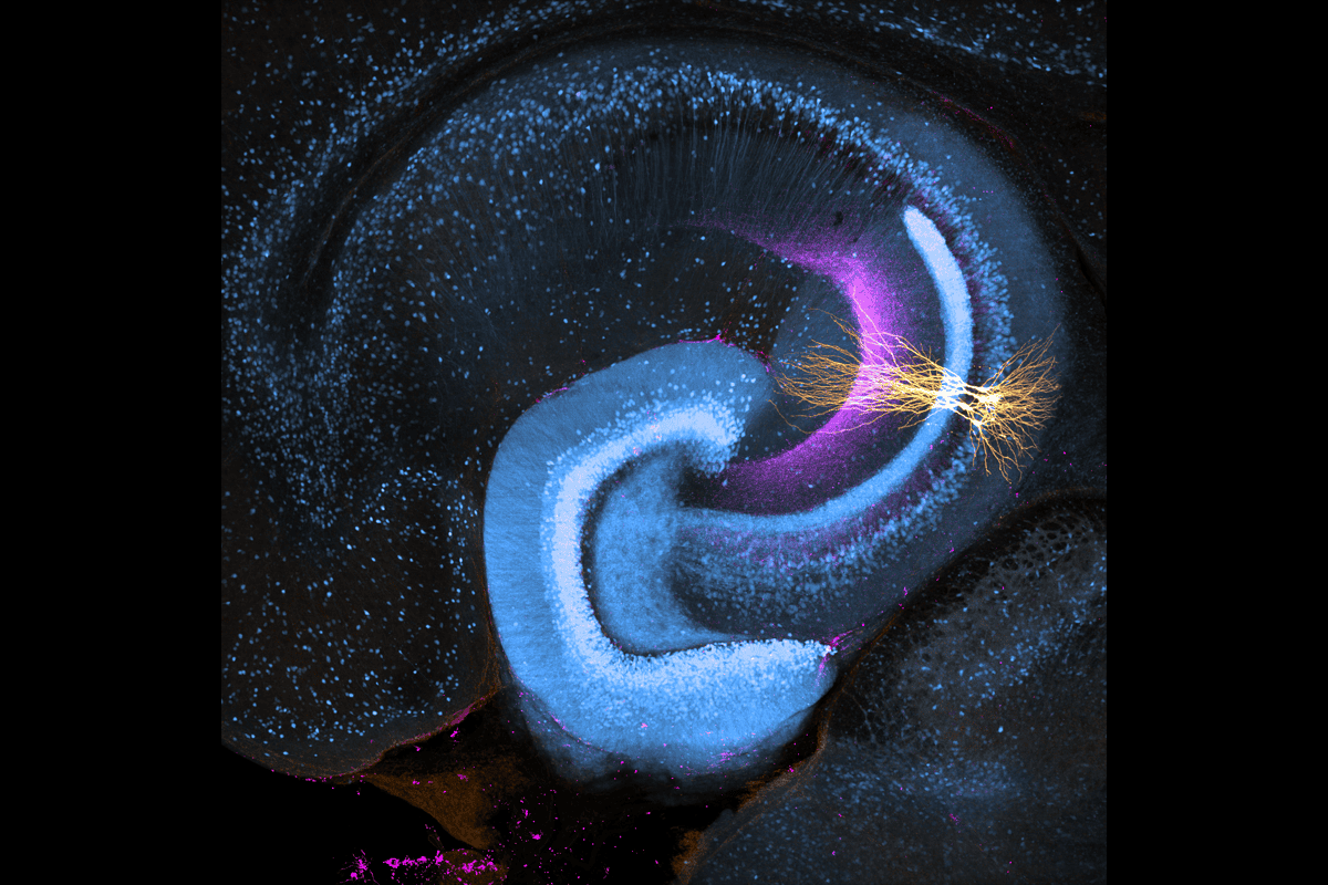
St18-expressing neurons.
Main target: The St18-expressing neurons exhibit an unusual projection pattern, primarily sending their axons (purple) to St18-negative neurons within the same hemisphere.

P

revious studies had documented differences in morphology, electrophysiology and connectivity among CA3 pyramidal neurons, Watson says, but the field lacked a unifying framework to explain how those differences fit together.

Watson’s team homed in on the ST18 expression distinction by analyzing publicly available single-cell RNA sequencing data from mice. They didn’t expect to see such a strong genetic distinction between CA3 cell types, Watson says. 

The ST18 expression also occurs deep in the CA1, the preprint shows. “I think the new component of this study is that these differences extend to the CA3 region as well,” says Attila Losonczy, professor of neuroscience at the University of Texas Southwestern Medical Center, who was not involved in the work. 

Additional experiments in mice engineered to express fluorescent ST18 protein revealed that the two groups of CA3 neurons also differ in where they send their connections. Superficial CA3 neurons follow the classical pattern, projecting broadly to other CA3 neurons and the CA1 region in both hemispheres.

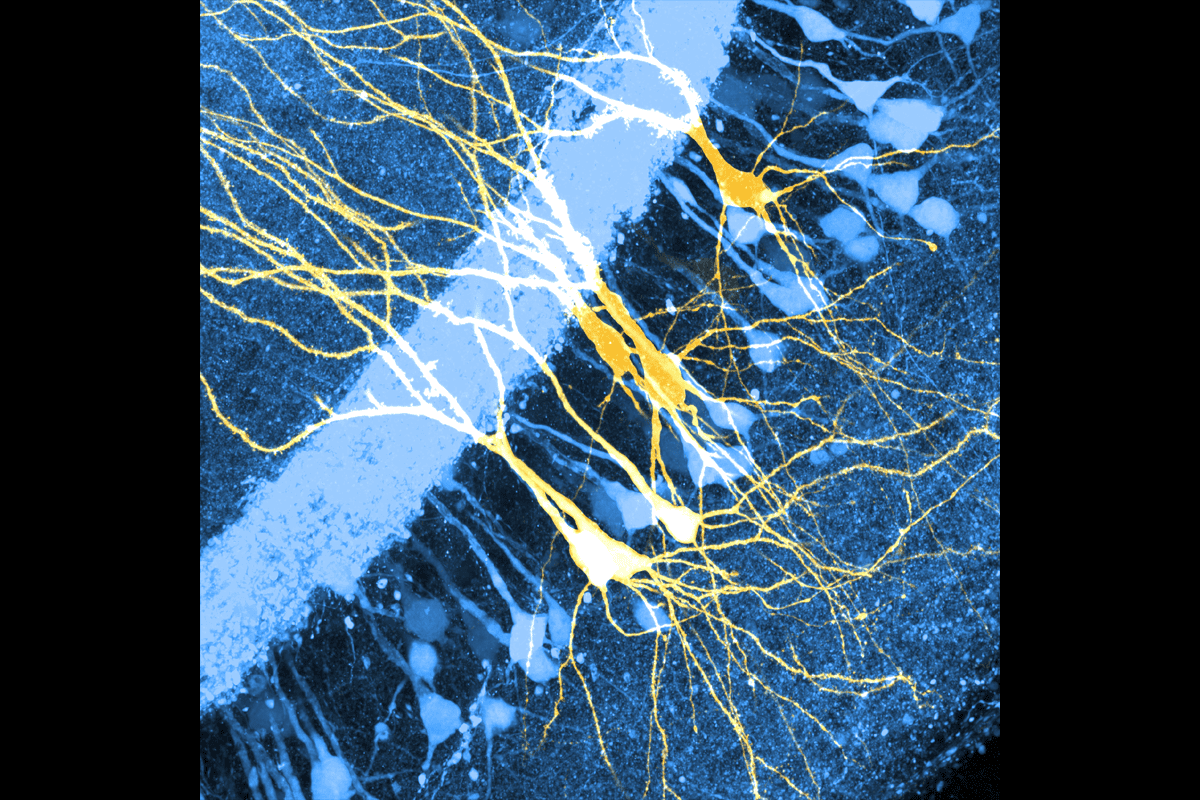
St18-positive neurons and St18-negative neurons.
Shape shift: The St18-positive neurons (blue) have simpler, less branched apical dendritic trees, whereas the ST-18-negative ones (yellow) have larger, more elaborate apical arbors.

Deep CA3 neurons, however, have about five times fewer connections to the CA1, and they project primarily within their same hemisphere. “They’re hemisphere specific, and controlling this memory network, it’s very weird and very cool,” Watson says. 

The deep cells also project to a narrow band on the distal apical dendrites, at the base of stratum radiatum of the entire CA3 region, and long-range projections from deep cells appear to influence superficial CA3 neurons through a combination of direct excitation and feedforward inhibition. “Locally, their connectivity seems to be different to what we have seen long range,” says study investigator Rebecca Morse Mora, a graduate student in Jonas’ lab.

The two sets of neurons show similar gene-expression differences in human and pig datasets. And they show similar morphological and connectivity differences in hippocampal samples from two people with epilepsy.

The cross-species comparison is one of the study’s strengths, Losonczy says—and “means that this computation must be preserved,” Buzsaki adds.

But the human data come from only a small number of cells and people, says Amelia J. Eisch, professor of neuroscience at the University of Pennsylvania, who was not involved in the work. So whether the deep network regulates the superficial one remains a proposed and not an established mechanism, she says.

The deep cells’ unusual projection pattern points to the next steps, Watson says: “What it means for brain function is the next question that is beyond what we have done.”

Sign up for our weekly newsletter.

Catch up on what you missed from our recent coverage, and get breaking news alerts.