A new functional MRI (fMRI) technique cuts down on scanning noise and motion-related artifacts, broadening the range of brain scans possible in awake and behaving animals, according to a new study.
“This is really groundbreaking work,” says Alessandro Gozzi, director of the Functional Neuroimaging Laboratory at the Istituto Italiano di Tecnologia, who was not involved in the study. One of the biggest challenges for scientists doing fMRI in awake animals has been conventional methods’ sensitivity to motion. The degree to which the new method reduces motion artifacts “is really remarkable,” Gozzi says. “The fact that it is silent is just a bonus to me.”
Because animals cannot be instructed to lie still for an fMRI scan, scientists typically anesthetize them first, which limits the types of experiments that can be carried out and raises questions about how sedatives might influence brain activity, says study investigator Yen-Yu Ian Shih, professor of neurology at the University of North Carolina at Chapel Hill.
Similarly, Shih says, stress and other factors may influence scans in awake animals that are acclimated to restraints, due to the noise levels, which can reach more than 100 decibels in conventional fMRI as the scanner sweeps back and forth and rapidly switches magnetic field gradients to acquire blood-oxygen-level dependent (BOLD) contrasts.
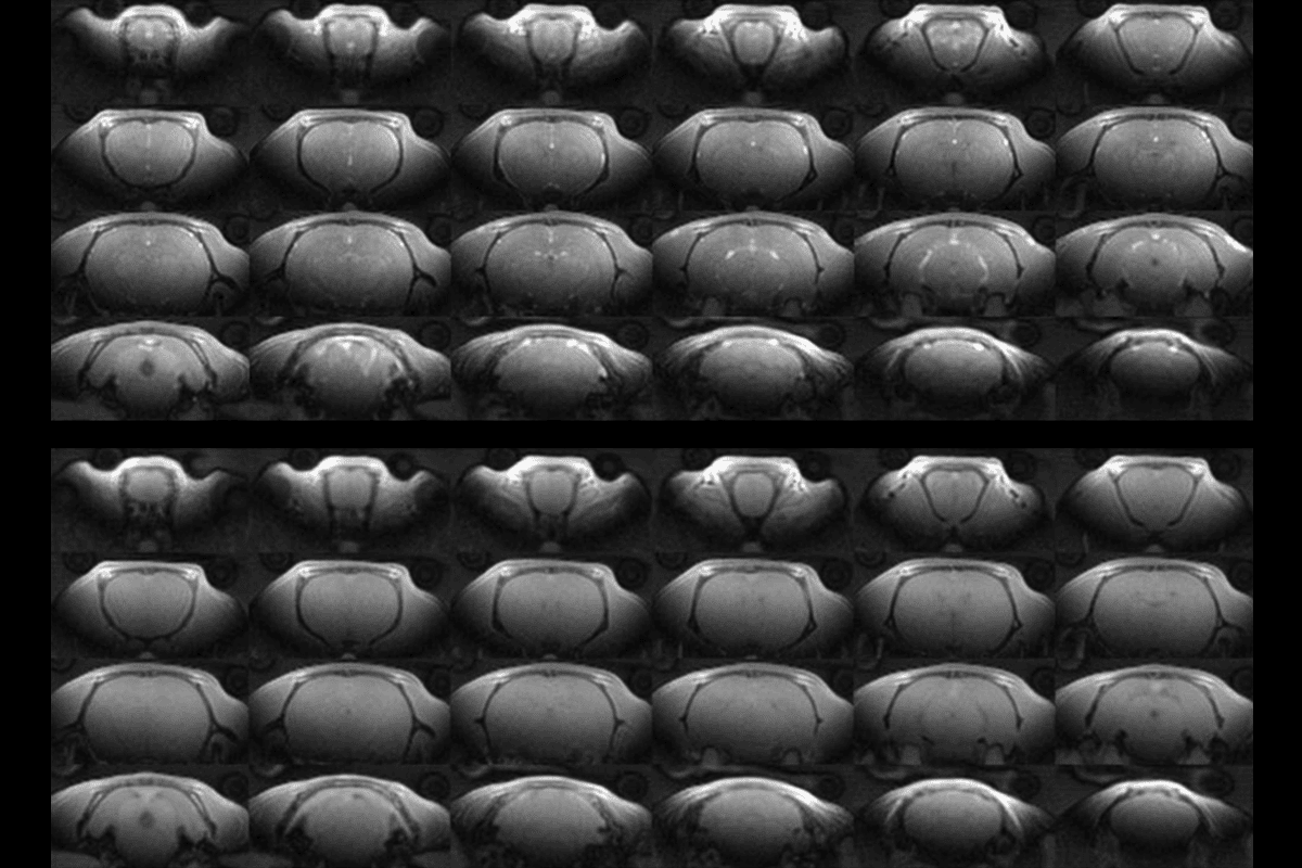
The new technique, dubbed “Steady-state On-the-Ramp Detection of INduction-decay with Oversampling” (or SORDINO, the word used to instruct musicians to mute their instruments), captures imaging data by rotating smoothly through space, akin to how the hands of a clock sweep across its face. This method keeps the magnetic field gradient nearly constant while also reducing the time between the radiofrequency pulses generated by the scanner and data acquisition to nearly zero. These findings were published last month in Nature Neuroscience.
This is a “very exciting advance,” particularly for animal studies, says Ravi Menon, professor of medical biophysics, medical imaging, neuroscience and psychiatry at the University of Western Ontario, who was not involved in this work. “They’ve combined a bunch of existing physical principles into a very clever application for functional MRI.”
And unlike the “direct imaging of neuronal activity” (DIANA) fMRI method that was the subject of intense scrutiny—and, eventually, a retraction—this technique is rooted in well-established physiology that underlies many MRI techniques, including BOLD-based fMRI, Menon adds.