Two competing sets of genetic instructions direct the brain’s cortical development in multiple species, a new study finds.
The program that guides the formation of the association cortex—which comprises regions that facilitate higher-order cognition, including abstract thought—activates around the frontotemporal poles of the developing cortex and spreads inward, according to the study, which analyzed spatial gene-expression data from mice, macaques, humans and other species. Soon after, sensory input from the thalamus kicks off a competing program that suppresses the development of the association areas in selected regions, carving out islands of the sensorimotor cortex.
The new work “provides a mechanistic step in understanding how [the sensorimotor-association] axis emerges,” says Daniel Margulies, research director at the National Centre for Scientific Research (CNRS), who was not involved in the study. It “also helps to ground that developmental trajectory in a way that gives us a framework for thinking about where it can go wrong.”
Humans devote a larger proportion of the cortex to association areas than other mammals do, but how this expansion evolved has been unclear. One influential explanation—the tethering hypothesis—proposed that primates decoupled the association cortex from the developmental constraints that shape sensory regions. As the primate cortex enlarged during evolution, sensory and motor regions remained relatively fixed, while the association cortex occupied the remaining territory.
The new study proposes a different mechanism—referred to as the multinodal induction-exclusion in network development (MIND) model. Rather than becoming untethered from primary sensorimotor areas, association areas actively compete with these regions to establish cortical terrain.
“This is an incredibly provocative and interesting idea,” says Fenna Krienen, assistant professor of neuroscience at Princeton University, who was not involved in the work but co-authored a 2013 paper outlining the tethering hypothesis. The model, and the findings it is based on, depart from the widespread notion that the development of the association cortex is delayed relative to sensory areas, she says.
The fact that multiple species have the same competing developmental programs suggests that primates did not evolve new mechanisms to expand the association cortex, says study investigator Nenad Sestan, professor of neuroscience at Yale University. Instead, evolution appears to have modified ancient developmental programs already present in the vertebrate brain, he says.
S
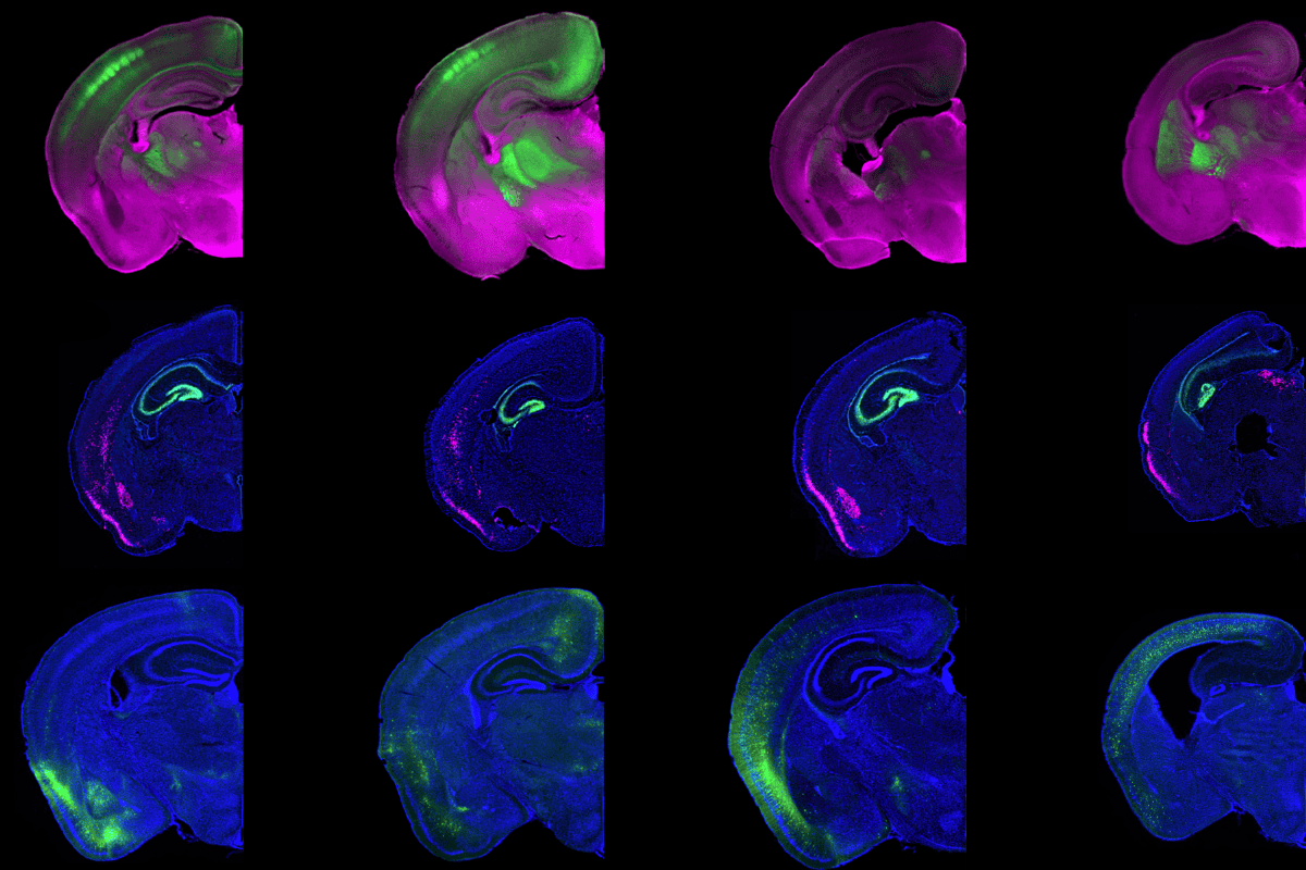
estan and his colleagues discovered the two competing developmental programs in cortical samples from fetal human and macaque brains of varying developmental stages. The pericentral program promotes the association cortex through genes involved in axon guidance and retinoic acid signaling. The central program specifies primary sensorimotor regions through a different set of genes. During development, the two programs become increasingly segregated as cortical regions are established, the study found.Evidence from two other species supports that model, the investigators showed. Mice lacking certain genes involved in cortical development—including the autism-linked genes SATB2 and ZBTB18—show reductions in sensorimotor areas and an expansion of the association cortex. Other mice, engineered to lack brain structures that connect the thalamus to the sensorimotor cortex, show a similar enlargement of association areas. And opossums, which have no primary motor cortex, show the association cortex in its place, the study found. “In my opinion, this was the key experiment of the paper,” Sestan says. “It tested the hypothesis in naturalistic evidence.”
The researchers also identified a potential molecular mechanism underlying this competition. The axon-guidance proteins SEMA7A and PLXNC1 are enriched in the primate sensorimotor and association cortex, respectively, and display complementary expression patterns across the developing cortex. When the team extracted cells from the two regions in mice and cultured them, they found that the neurons repelled each other. But cocultured cells taken from rodents that lacked either SEMA7A or PLXNC1 showed no such repulsion. The same molecular markers were found in mice, opossums and chickens, indicating that this organizational principle evolved before the emergence of primates.
That inverse expression of sensorimotor and association genes across multiple species “opens new doors in comparative evolutionary neuroscience,” says Valerie Sydnor, a postdoctoral scholar at the University of Pittsburgh, who was not involved in the study. “This underscores how there is much to learn about the human brain through cross-species approaches.”
R
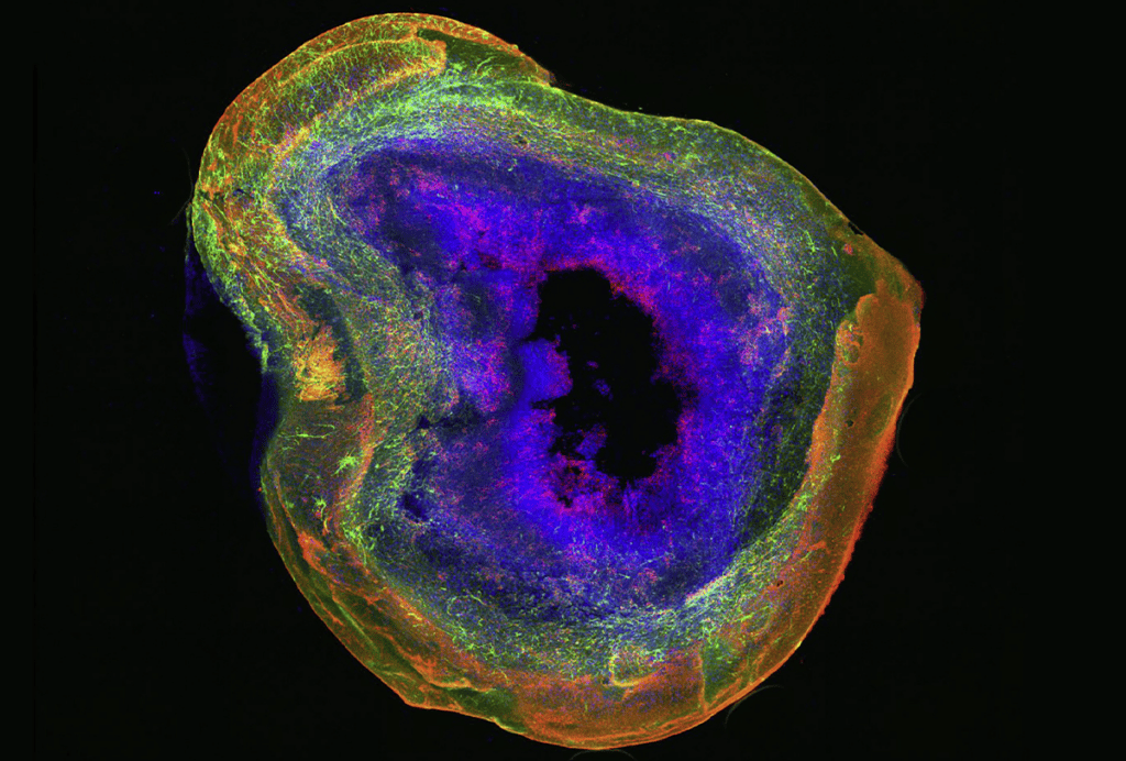
etinoic acid appears to promote the formation of the association cortex, the study also found. Human cortical organoids treated with the molecule increase their expression of PLXNC1.Autism-linked genes were enriched in both developmental programs, suggesting that altered regulation of these pathways could shift the balance between the sensorimotor and association areas during development. Such a mechanism could explain the altered organization of these regions observed in autistic people in neuroimaging studies, says study investigator Jeremiah Tsyporin, a postdoctoral associate at Yale University.
The findings were published earlier this month in Nature.
The work “provides a new vocabulary for asking how cortical hierarchy emerges” that could ultimately extend beyond the cortex, says Zoltán Molnár, professor of developmental neuroscience at the University of Oxford, who was not involved in the work. But the model remains incomplete without accounting for the thalamus, whose development likely influences the balance between the sensorimotor and association territories, he says.
Though the study investigators don’t plan to incorporate the thalamus into the model, their findings suggest that similar mechanisms are involved, Sestan says. SEMA7A is enriched in first-order thalamic nuclei—neuronal clusters that relay signals to sensorimotor areas—whereas PLXNC1 expression is strongest in parts of the thalamus that project to the association cortices, the study found.
The study investigators next plan to investigate how the competition for cortical territory is affected in animal models of autism and other complex neurobiological disorders, Tsyporin says.