LISTEN TO THIS STORY:
0:00
/
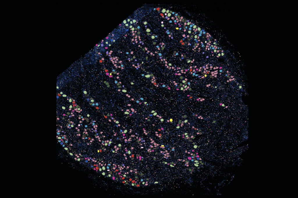
The spatial arrangement of neurons in the locus coeruleus of mice corresponds with the cells’ targets across the brain, according to a new study.
Neurons in the locus coeruleus, which provides norepinephrine to the rest of the brain and spinal cord, are more spatially and functionally diverse than previously thought, a new preprint finds. The work reveals how such a small structure located deep in the brainstem can influence a range of functions in multiple brain regions.
Locus coeruleus neurons show gene expression variations that track with differences in the cells’ shape and projection targets, the study found. And neurons that occupy opposite ends of the structure respond differently to the rewards mice receive during a learning task, suggesting that the neurons facilitate learning in distinct ways.
“This is the bread-and-butter work that the locus coeruleus field needed,” says Nelson Totah, associate professor of neurophysiology and pharmacology at the University of Helsinki, who was not involved in the study. “What they did here was not ask flashy questions [but] answer fundamental questions about this evolutionarily ancient nucleus, so I’m really glad to see this work.”
The locus coeruleus—which translates from Latin to “blue spot”—is named for the blue pigmented cells that synthesize norepinephrine. The structure was long thought to consist of homogeneous neurons that secrete norepinephrine in synchrony. But over the past two decades it has become increasingly clear that the region is structurally and functionally heterogeneous: It has two distinct neuronal subtypes that fire asynchronously and drive opposite behaviors in rats, according to papers published in 2018 and 2017, respectively.
The new findings suggest that the structure’s neurons are even more diverse and follow a precise organizational pattern: From one end of the region to the other, neurons show a spatial gradient in gene expression differences that map onto variations in the cells’ morphology, electrical activity and target regions, the new study found.
“This study goes to the next level in terms of demonstrating the spatial organization of heterogeneity at multiple levels,” says Rebecca Jordan, principal investigator of the Prediction & Plasticity Lab at the University of Edinburgh, who was not involved in the work.
N
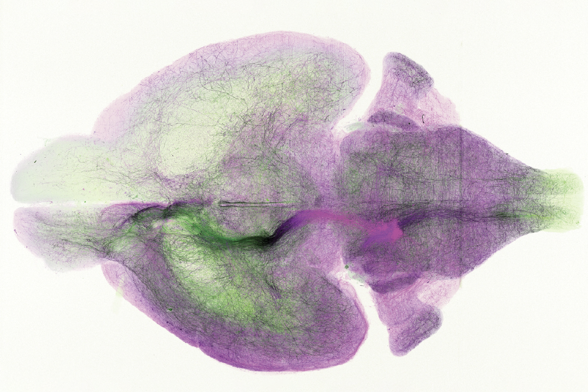
euronal labeling in the mouse locus coeruleus revealed that individual neurons tend to target specific brain areas that collectively project to almost the entire central nervous system, the researchers found. The labeled neurons innervate wide swaths of brain tissue, suggesting that cells projecting from the region have a large area of influence.Some labeled neurons had axons measuring up to 73 centimeters long. “As far as we know, it is the longest neuron that has been reconstructed in a mouse,” says study investigator Jeremiah Cohen, principal scientist at the Allen Institute for Neural Dynamics. “[Who knows] what sort of length you or I have in our heads?”
Across the locus coeruleus—from dorsal to ventral regions—Cohen and his colleagues detected smooth variations in gene expression. Depending on their position in that gradient, neurons project to distinct target areas, they found. For instance, dorsal and ventral neurons—which represent the extreme ends of the gene expression gradient—send projections to the cortex and spinal cord, respectively. Neurons in the center of the structure targeting the cerebellum show an intermediate pattern of gene expression.
That “gene expression profiling gives us a potential handle on trying to record and manipulate in a more cell-type-specific manner,” says Robert Froemke, professor of neuroscience and otolaryngology at New York University School of Medicine, who was not involved in the study.
Spatially distinct neurons also show different patterns of activity. Dorsal neurons are highly active when mice change their behavior in a learning task or when they receive an unexpected reward, the researchers found. Because dorsal neurons project to the frontal cortex, norepinephrine released by those cells could serve as a learning signal, Cohen says.
By contrast, ventral neurons respond strongly whenever the mouse repeats a behavior or when it doesn’t receive a reward. The findings were posted on bioRxiv last month.
That diversity in reward responses, which appears to correlate with projection targets, suggests “that the locus coeruleus can, in principle, send tailored information to different brain areas depending on their function,” Jordan says.
Cohen and his team next plan to determine how cortical circuits harness the neurotransmitter to drive learning and its interactions with other neuromodulatory systems, he says. So far, little is known about how norepinephrine interacts with dopamine and serotonin to influence mammalian brain circuits, Cohen says. “I would love to have a real theory of what these neuromodulators do to circuits and behavior.”