The human brain forms trillions of synapses using only a few thousand cell-surface proteins to guide the process. How can distinct functional circuits form when there are far more neurons than molecules to guide them?
New findings in mice provide an answer to this numerical puzzle: Across the brain, neurons repeatedly reuse the same pair of adhesion molecules to assemble distinct neural circuits, according to three studies published last month in Current Biology. The molecules—teneurin-3 (TEN3) and latrophilin-2 (LPHN2)—help direct axons to their targets using a common set of wiring rules, the studies show.
The molecules are especially important in sensory regions and in the formation of body maps, the new work suggests. The findings could help to uncover pathways that lead to sensory sensitivity in people with autism or related conditions.
“It seems the brain is reusing these molecular cues where it can,” says Daniel Pederick, assistant professor of neuroscience at Johns Hopkins University, who worked on all three studies. Connections from distinct brain areas—such as the visual system and the brainstem—“are never going to come into contact so won’t interfere with each other.”
The work is a “nice extension of ideas that are dominant in the field,” says Peter Robin Hiesinger, professor of neurobiology at Freie Universität Berlin, who was not involved in any of the new studies. “Even though it’s just two molecules, their context-dependent function in different neurons and brain regions is likely multitude.”
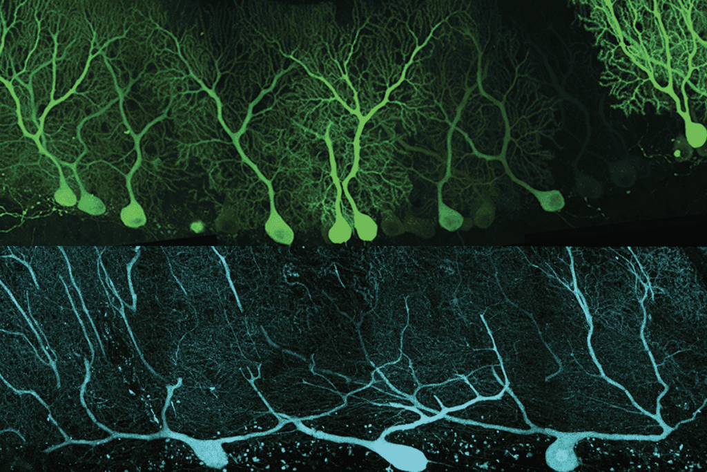
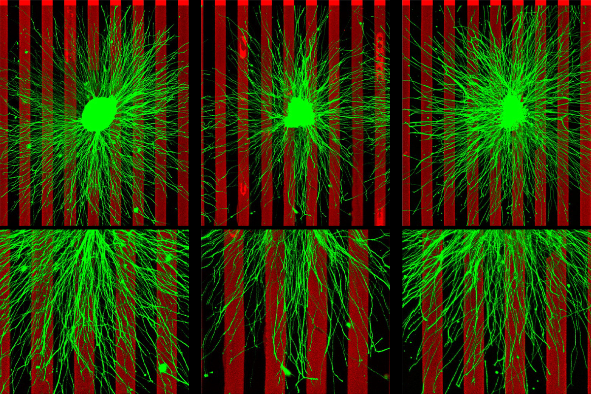
TEN3 and LPHN2 guide neuronal targets using a combination of attraction and repulsion, according to a 2021 study. In specific circuits of the mouse hippocampus, TEN3 stabilizes connections between axon terminals and dendrites that also express the molecule, whereas repulsive interactions between TEN3 and LPHN2 prevent axons from forming off-target connections, that study found.
T
hat “push-pull” system is active across the mouse central nervous system, including in the extended hippocampus, visual and auditory systems, cerebellum, basal ganglia and spinal cord, the new studies found. In each area, TEN3 and LPHN2 form opposing expression patterns that direct axons to their targets. Mice lacking these molecules develop unusually diffuse projections with misplaced connections, according to one of the studies.Though both attractive and repulsive signals are essential, “the relative importance of [those forces] depends on whether the developing axon first encounters the attractive or repulsive field,” says Liqun Luo, professor of biology at Stanford University, who has been working on this puzzle since 1999 and is an investigator on the three new studies. If a growing axon passes through the wrong region, repulsion becomes especially important, he says. “It tells them, ‘Hey, this is not your target; keep going,’ until it reaches the attractive field.”
And those expression patterns mirror the region’s functional organization. In the auditory system, for example, the proteins display opposing gradients that correspond to the spatial mapping of sound frequencies: LPHN2 is most strongly expressed in neurons that process high-frequency sounds, whereas TEN3 levels peak in neurons tuned to low frequencies.
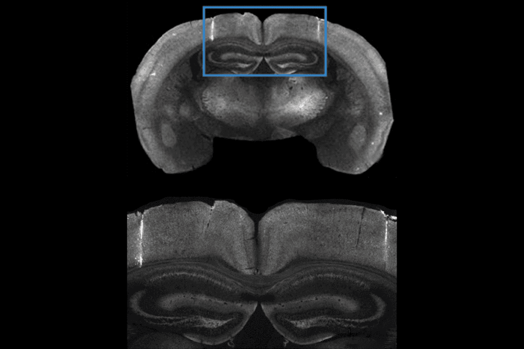
Disrupting those patterns also distorts the brain’s body maps. Mice lacking TEN3 in neurons of the dorsal horn—a region of the spinal cord that receives sensory signals from the body—formed representations of the limbs that were compressed or expanded compared with typical body maps, the researchers found by injecting tracers into different parts of the mice’s limbs and detecting their presence in the spinal cord. The engineered mice also spent more time licking the wrong location following a painful injection in one of their hindpaws than did their wildtype counterparts, the study found.
Until now, how the brain assembles precise body maps remained a mystery, says Claude Desplan, professor of biology and neural science at New York University, who was not involved in the work. The new work is an “important advance” that reveals how neurons connect to establish a rough map, though it’s unclear how those maps are refined, he says.
Although TEN3 and LPHN2 help to build initial proto-maps, they are likely refined through neuronal activity, says study investigator Artur Kania, lab director at the Montreal Clinical Research Institute. Such processes may allow body maps to adapt to individual experiences—for example, by reshaping the neural territory devoted to sound processing in deaf people or trained musicians.
A more complete understanding of how sensory maps are assembled could help explain why some people with autism or related conditions struggle to process sensory information, Pederick says. He and his team next plan to investigate whether auditory maps are altered in mouse models of autism, he says. Because many genes strongly associated with autism encode transcription factors, they will examine whether variations in those genes disrupt expression of TEN3 and LPHN2, and how that impacts auditory processing, he says.