Neuroscience textbooks have long cast mitochondria as pure neuronal powerhouses: These bean-shaped organelles just crank out a cell’s energy. That picture, however, is starting to look incomplete.
Mitochondria do far more than fuel neurons, a growing body of research suggests. They also appear to help synapses communicate, regulate neurotransmitter release and shape social behavior. Mitochondrial function has also been tied to autism and related neurodevelopmental conditions, though that link remains debated.
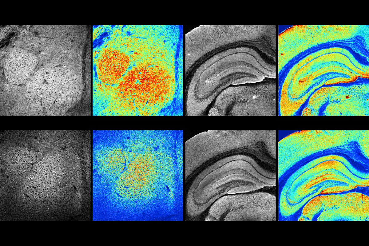
Even memory formation may lean on these tiny, double-membraned structures, according to a study published in Nature Metabolism in February. Increasing mitochondrial metabolism boosted long-term memory in both fruit flies and mice.
Mitochondria are “not just permissive but also instructive,” says Ezgi Hacisuleyman, assistant professor of molecular medicine at the Herbert Wertheim UF Scripps Institute for Biomedical Innovation & Technology, who was not involved in the February study. Her unpublished results show that mitochondrial proteins are translated near active synapses, for example.
Over the past decade, work from Hacisuleyman and others has fast expanded the repertoire of mitochondria in the brain. Taken together, she adds, the findings put mitochondria “more in the center of how we think about brain function and memory.”
M
“Synapses are energy-hungry compartments,” says Vidhya Rangaraju, who leads a research group at the Max Planck Florida Institute for Neuroscience. “So they have to have local power plants.” Dendritic mitochondria can form local, stable structures—about 30 micrometers long—to fuel protein translation during synaptic remodeling, her work has shown.
These local mitochondria typically respond to calcium: When neurons fire, calcium ions enter nearby mitochondria and prompt them to rapidly increase energy production. An increase in mitochondrial calcium signaling also boosts metabolism in memory circuits and, as a result, improves brain function, according to the February study.
In addition, lowering levels of a protein called LETM1, which would otherwise move calcium out of mitochondria, pushes the organelles into a more active metabolic state. This metabolic boost helped flies and mice form lasting memories that untreated animals did not: Flies remembered an odor paired with a mild shock one day after a single training session, and mice remembered an odor associated with lithium chloride exposure—making them temporarily feel sick—several days later.
“Our results show that energy not only enables brain function, but it may also govern brain function,” says study investigator Jaime de Juan-Sanz, who leads a research group at the Paris Brain Institute. Some neural circuits may be constrained by how much energy they can generate, he says, so added metabolic capacity could also improve cognitive performance.
A
In flies, for example, mitochondrial ROS help drive long-lasting synaptic remodeling early during development, a 2025 study suggests. Lowered mitochondrial ROS signaling led to unusual structural plasticity at the neuromuscular junction, where a motor neuron signals to a muscle cell. At the same synapse, increasing ROS in motor neurons—but not in muscle cells—helped preserve communication between nerve and muscle, another 2025 study found.
“ROS can be used to signal important things for the cell,” says Andrew Frank, professor of anatomy and cell biology at the University of Iowa’s Carver College of Medicine, who worked on the latter 2025 study. “If you just shut it off, you may be interfering with the natural signaling process,” he says.
The findings support the idea that instead of just fueling growth, mitochondria may also instruct when and how synapses change during sensitive developmental windows, Frank says.
On the other side of the lifespan, mitochondrial function seems to support a protective response neurons activate as flies age, increasing levels of proteins involved in neurotransmitter release at synapses. Reducing mitochondrial function through genetic manipulation made this age-related increase stronger, whereas improving it dampened the response, a third 2025 study showed.
“While they are examining different synapses and different mitochondrial manipulations than we did, some of their findings match our presynaptic results nicely,” Frank says, and they also point to a shared theme: When mitochondrial signaling is disrupted, synapses respond by increasing specific proteins at sites of neurotransmitter release.
ROS signaling may even help explain why neurons fire when they seem to have nothing to say. Some self-generated firing may prevent ROS buildup and thereby help neurons maintain a metabolic balance when ATP consumption falls too low, according to a 2023 computational model.
Neurons must constantly manage energy and oxidative stress, which can lead to cellular toxicity, so “it seems fundamentally important to make these spontaneous [spikes],” says André Fenton, professor of neural science at New York University, who was not involved in the work. The idea has some indirect support from work in fruit flies showing that when mitochondrial redox signals change, sleep-control neurons alter how readily they fire.
Testing whether silencing neurons causes ROS to build up is experimentally difficult, says Tim Vogels, professor of computational neuroscience and neurotheory at the Institute of Science and Technology Austria, who helped develop the 2023 model. But “spontaneous activity may be less like noise and more like a pressure-release valve for the cell,” he says.
B
For example, rats that show anxiety-like behavior are more likely to lose social competitions, and, researchers found in 2015, this disadvantage is linked to weaker mitochondrial function in the nucleus accumbens—a region involved in motivation. Reducing mitochondrial function there made typical rats lose competitions over new territory, whereas boosting mitochondrial metabolism helped the ones with anxiety-like behavior avoid becoming subordinate.
“That was one of the first works that showed that manipulating mitochondrial function directly influences behavioral outcomes,” says study investigator Fiona Hollis, assistant professor of pharmacology, physiology and neuroscience at the University of South Carolina.
Mitochondria have since been linked to other stress-related behaviors. In mice, chronic social defeat stress damaged the organelles in the amygdala and triggered their depletion in a circuit that normally helps buffer anxiety, a 2021 study found. The same type of stress altered contacts between mitochondria and the endoplasmic reticulum in hippocampal microglia, contributing to mitochondrial damage, inflammatory signaling and depression-like behavior, another study showed.
The fact that some animals are more or less resilient to stress may be due to the wide variation of mitochondrial activity across the mouse brain and its correlation with anxiety-like behavior and social avoidance, says Martin Picard, associate professor of behavioral medicine at Columbia University, describing his recent work.
Such naturally varying mitochondrial molecules or functions might also contribute to cognitive differences across individuals, says Carmen Sandi, professor of neuroscience at the Ecole Polytechnique Federale de Lausanne, and help explain the February findings that link mitochondria to memory.
The organelles may even help regulate sleep. In flies, mitochondria in sleep-control neurons broke into smaller pieces and were recycled after sleep loss, and altering this process changed how much the animals slept, 2025 findings suggest.
The mechanisms through which mitochondria may shape cognition and behavior are still unclear, Sandi says. But, she adds, “we should really stop thinking only about energy. … The emerging view is that mitochondria could be important for brain health.”