Individual structural MRI studies fail to identify reproducible brain signatures for autism and other neuropsychiatric conditions, a new study finds.
Brain scans of people with autism, depression and bipolar disorder rarely show consistent differences in cortical thickness or grey matter volume across independent studies, according to the analysis. The study suggests that these structural measures are unlikely to provide reliable biomarkers of those conditions and that different approaches are needed.
“The findings are really a wake-up call. We’ve all been operating under the assumption that if we keep doing these studies enough, eventually the noise will wash out and we will converge on some consensus of what the brain changes in a particular disorder are,” says study investigator Alex Fornito, professor of psychology at Monash University. “Our findings suggest if we keep doing that business as usual, that’s not going to happen.”
Structural MRI studies have long produced conflicting results. Some have identified greater cortical thickness of select brain regions in people with autism than those without the condition, while others have reported the opposite. Until now, it was unclear whether such discrepancies reflected differences in study design and analysis, or whether structural brain signatures for these conditions do not exist.
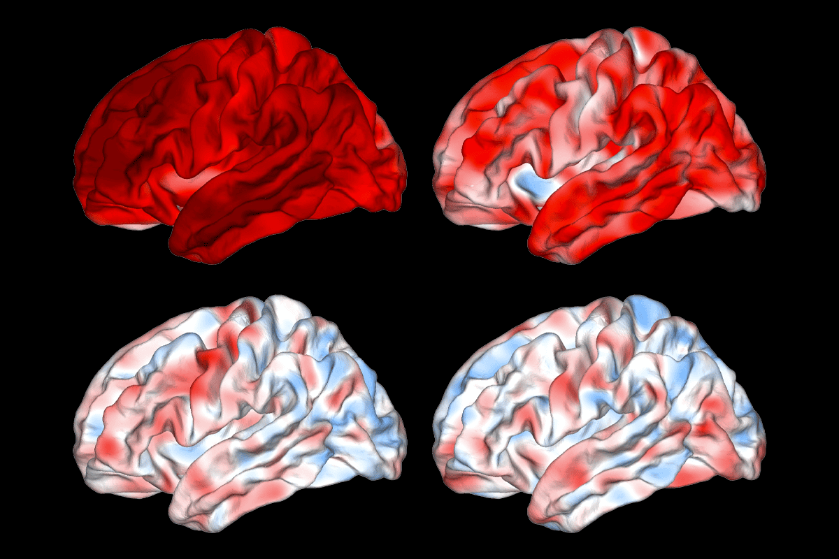
To address that question, Fornito and his colleagues analyzed MRI scans from thousands of people with neuropsychiatric conditions—including depression, schizophrenia, schizoaffective disorder, autism and bipolar disorder—as well as from people with Alzheimer’s disease. They calculated cortical thickness or grey matter volume using the same analysis pipeline across all datasets and then compared how consistently brain changes were reproduced between independent study sites.
Unlike Alzheimer’s disease, which showed a robust and reproducible brain signature, the neuropsychiatric conditions initially showed little agreement across studies. Accounting for demographic and technical factors, including age, sex and scanner differences, failed to explain the inconsistency, the study found.
“We need to temper our expectations by acknowledging that there is only so much that MRI and other neuroimaging techniques can tell us,” says Joshua Roffman, associate professor of psychiatry at Harvard Medical School, who was not involved in the study. “For example, measurement of cortical thickness—one of the main imaging markers described in this study—comes along with an intrinsic amount of measurement error, even under ideal conditions.”
U
sing mathematical models to simulate larger studies, the team found that for schizophrenia, reproducibility improved as sample sizes increased, reaching the same level as for Alzheimer’s disease with simulated cohorts of more than 200 participants. But the same pattern did not emerge for autism, depression, schizoaffective disorder or bipolar disorder—though datasets exceeding 150 participants were limited for most of these conditions.The findings were published last month in Nature Neuroscience.
That difference could reflect the more stringent criteria used in diagnosing schizophrenia, Fornito says. For example, a schizophrenia diagnosis requires a consistent pattern of symptoms for six months. In contrast, diagnoses of autism, bipolar disorder and depression apply to people with broader ranges of clinical presentations, he says.
Neuropsychiatric diagnoses are thought to correspond to distinct brain changes, says Louise Mewton, associate professor of public health at the University of Sydney, who was not involved in the work. But “across this study and many others, we are not seeing evidence of discrete categories of mental disorders.”
Alternative frameworks that classify people according to trait profiles—such as the Hierarchical Taxonomy of Psychopathology (HiTOP) model—might help researchers uncover more reliable structural biomarkers, says study investigator Trang Cao, a research fellow at Monash University. Repeating the analysis using cohorts characterized in this way could reveal more consistent brain signatures, she says.
But imaging alone is unlikely to provide the answers, says Maria Di Biase, associate professor of psychiatry and neuroscience at the University of Melbourne, who was not involved in the study. Rather than serving as a “standalone diagnostic tool, structural MRI will likely be most informative when integrated with genetics, molecular biology and longitudinal clinical data.”
Large collaborative efforts are already moving in that direction. The ENIGMA consortium pools brain imaging data from 43 countries and combines it with genetic and epigenetic information to identify subtle biological signatures that would be impossible to detect in smaller studies. That type of approach is “really going to help us pick apart which brain changes are robust [from] those which aren’t,” Fornito says.
Next, the team plans to repeat the analysis using larger unsimulated datasets to determine whether reproducibility improves as sample sizes increase, Cao says. Rather than focusing solely on regional measures such as cortical thickness and grey matter volume, they also plan to investigate whole-brain anatomical patterns to see if these provide reproducible signatures of neuropsychiatric conditions, she says.