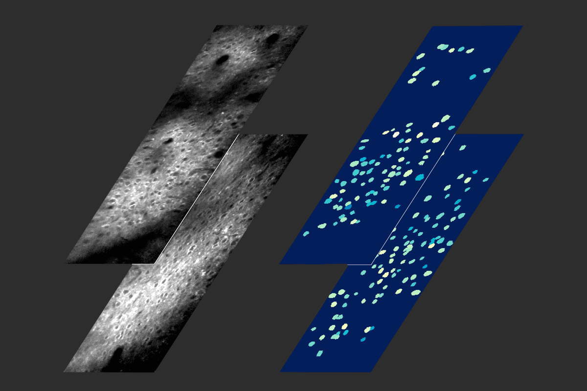
Dual view: Simultaneous imaging of layer 2/3 (top) and layer 4 (bottom) neurons of the mouse posterior parietal cortex reveals that the latter activate first after whisker stimulation.
Add us as a Preferred Source on Google

Optimized two-photon microscopy enables voltage imaging at multiple depths

The tool could reveal how information flows within and between cortical layers during neural processing.

By Simon Makin
11 August 2026 | 4 min read

A new optical imaging platform makes it possible to scan the activity of neurons across wider areas and deeper brain tissue, and even at different depths simultaneously. The platform is described in a paper published last month in Nature Methods.

“This is a highly optimized system for two-photon voltage imaging,” says Adam Ezra Cohen, professor of chemical biology and physics at Harvard University, who was not involved in the work.

Calcium indicators, which flash as calcium flows into neurons after they fire, have been the workhorse of optical brain imaging for over a decade. But calcium is a proxy for what scientists are really interested in—electrical activity—and it changes much more slowly, obscuring fine timing information.

Genetically encoded voltage indicators (GEVIs) track neural activity directly, but, until recently, only over small areas, measuring up to about 50 × 250 μm and typically involving around 10 neurons. GEVIs also sit in membranes, making them harder to resolve, and the signals they produce are weaker and last only around a millisecond. 

The tissue-scanning lasers in traditional two-photon microscopes can only move so fast. “You’re measuring one point at a time, so you have to scan very fast, typically 1,000 frames a second, or faster,” to image GEVIs, Cohen says, which “is exceptionally hard to do with a point scanning device.”

The microscopes also need to deliver as much energy as possible without heating tissue. “You have to walk this tightrope, where you put enough light into the brain to get signal out, without frying the brain,” says Jerry Chen, associate professor of biology at Boston University, who was not involved in the study.

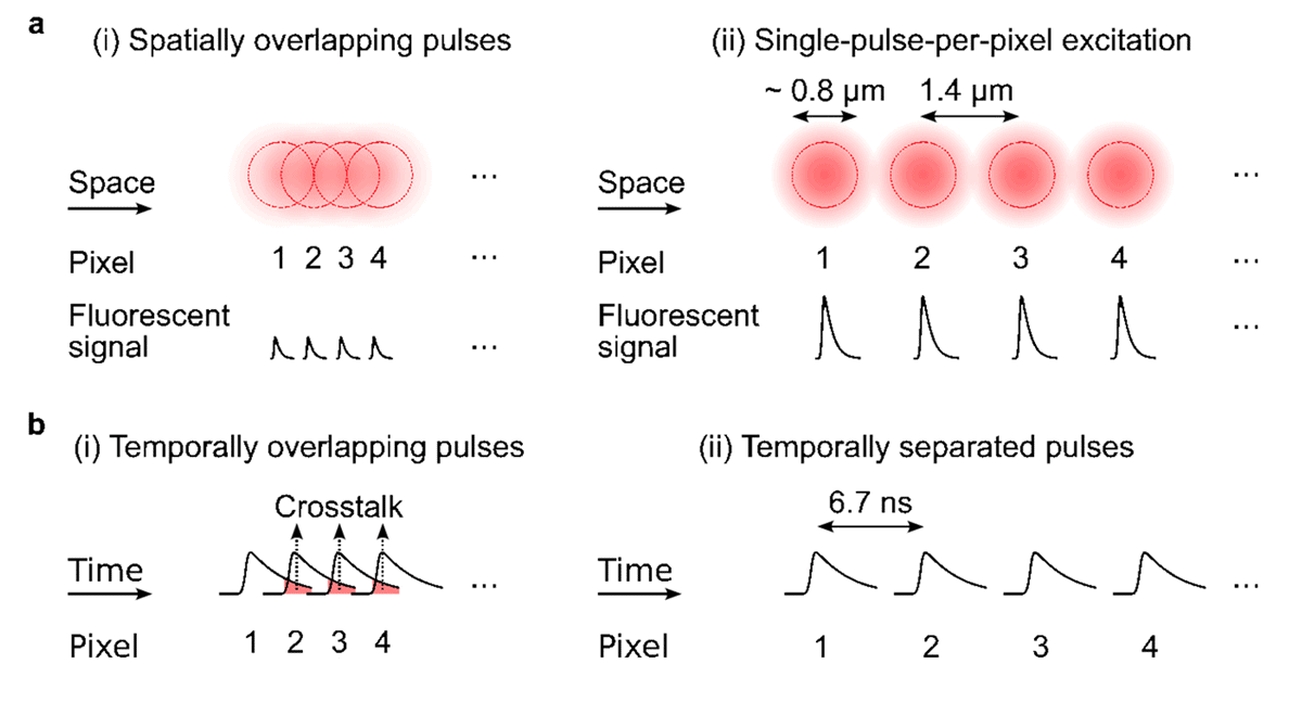
Adding to the complexity, if you place scan points too far apart, you can’t resolve small structures, but if they are too close together, you waste energy. “To optimize energy use, you want one pulse per pixel,” says study investigator Alipasha Vaziri, professor of neurosciences and behavior at Rockefeller University. Exciting the sensors repeatedly before they’ve settled back down can also cause them to burn out, a process known as photobleaching.

The new platform, called FlatMux, optimizes all these dimensions. “To maximize the population you can image, you need to think very strategically about how to maximize efficiency in terms of energy, time and space,” Vaziri says.
 

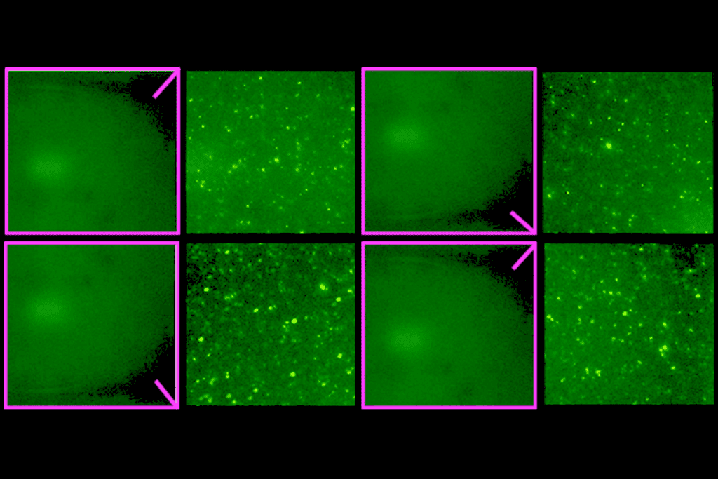
Research image of fluorescence signals.
Optimaxxing imaging: Optimal spatial placement of laser pulses results in higher fluorescence signal for the same power (top). A delay of 6.7 nanoseconds between pulses allows the fluorescent signal to decay, reducing crosstalk while also optimizing use of temporal resources and pixel acquisition rate (bottom).

T

he system involves arrangements of mirrors that split a laser beam into 14 “light beads.” A beam splitter doubles this to 28. “They split their laser into an arrayed comb of spots, then scan those [light beads] through the sample in parallel, so you can measure many more points at a time,” Cohen says. 

The mirrors produce slight delays between beads, making it possible to place excitation precisely and increasing the area that can be scanned—up to 590 × 400 microns, and in one case recording from 180 neurons simultaneously. The team also demonstrated a deep mode, recording from neurons 500 microns within the brain, and a rapid mode, capable of 2 kilohertz frame rates.

A similar approach, which also splits the laser and was published in December, solved many of the same problems, but “the main advance in the newest work from Vaziri is that the locations of the pulses can be more easily reconfigured,” Cohen says. This versatility is particularly useful for scanning two planes simultaneously.

Vaziri’s team demonstrated this multilevel scanning on mice while their whiskers were stimulated. “We put one plane in layer 2/3 and another in layer 4 of the mouse cortex,” Vaziri says. “By imaging these two planes, we showed that neurons in layer 4 were activated first, then layer 2/3 neurons.”

Neuroscientists could use this to trace how information flows, both “within and between cortical layers during sensory processing, or other cortical computations,” Cohen says.

Another configuration maximized sensitivity, making it possible to detect changes below neurons’ firing threshold. “Subthreshold activity is important because it gives information about connected neurons,” Vaziri says. “If you imagine combining this with techniques such as optogenetics, you move towards an optical approach for circuit mapping.”

The platform’s flexibility should enable the team to incorporate future advances in voltage indicators, which are constantly improving. “That was in our minds from the outset, to take advantage of ongoing improvements,” Vaziri says.

Going forward, however, the “cost and complexity [of the platform] are a challenge that will have to be dealt with if these are going to be widely used,” Cohen says.

Sign up for our weekly newsletter.

Catch up on what you missed from our recent coverage, and get breaking news alerts.