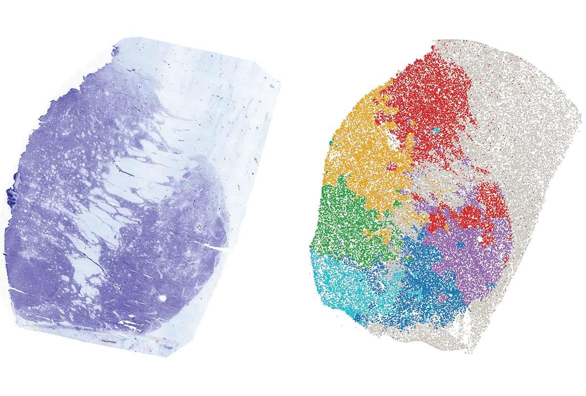
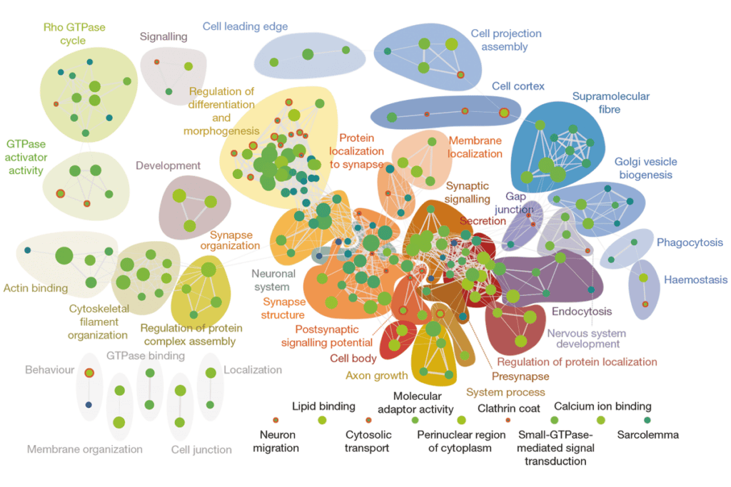
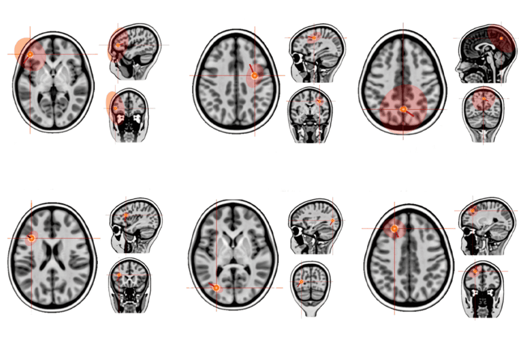
The basal ganglia comprises 61 consensus cell groups, some of which are noncanonical subtypes not previously characterized in detail, according to a single-nuclei, cross-species atlas published today in a package of 28 papers by the Cell Press family of journals. The atlas surveys 1.8 million nuclei from the basal ganglia of humans, macaques and marmosets.
“If you buy a kit to put something together, the first thing you take out is the parts list to make sure that everything’s there, and here we have that parts list [for the basal ganglia],” says Peter Strick, professor of neurobiology at the University of Pittsburgh, who was not involved with the effort. “There’s so much that will be built on top of this.”
The work is the product of the BRAIN Initiative Cell Atlas Network (BICAN), a consortium launched in 2022 to create high-resolution and comprehensive cell atlases, specifically of the human and nonhuman primate brains. After months of meetings and discussions, the group arrived at a common taxonomy of cell types in the basal ganglia based on gene expression, spatial and electrophysiology data.
This consensus set the stage to have a common language across all of the labs, says BICAN member Evan Macosko, associate professor of psychiatry and neurobiology at Harvard Medical School and a core member of the Broad Institute of MIT and Harvard. “If we don’t have a uniform naming system and understanding, then it just makes communication really difficult,” he says.
Leveraging this shared understanding, researchers studied gene expression, epigenetic and histone profiling, genome folding, and neuronal shape and electrical properties in the developing and adult basal ganglia. The atlas also provides fine-grained spatial transcriptomic information, revealing a stereotyped molecular architecture of regions such as the striatum, one of the studies shows.
“It wasn’t until we could actually map it to their spatial context that you can really appreciate that there is this spatial logic to how these cell types are changing across evolution,” says Fenna Krienen, assistant professor of neuroscience at the Princeton Neuroscience Institute, who worked on the project. “And sometimes it upends our classical views of how distinct different cell types are.”
The common taxonomy and precise molecular and spatial characterizations open the door for researchers in the field to find new biological insights or produce better tools, akin to the Human Genome Project, says John Ngai, BRAIN Initiative director. “Researchers do not have to go through this whole thing by themselves,” he says. Instead, they can use the atlas as a reference without having to perform all the analyses.
Some of the data are available on the BICAN portal, described in one of the papers, or featured on the institutional websites of BICAN members. Other studies in the package include resources, tools and frameworks that facilitate the integration, scaling and analysis of multi-omic data. (See a list of all the studies.)
“It’s a new phase of BICAN,” says Hongkui Zeng, director of the Allen Institute for Brain Science, who worked on the project. But it may also be the final phase, she adds. Although the ultimate goal is to expand and create an atlas of the entire primate brain, the BICAN project, which is funded by the U.S. National Institutes of Health, is slated to run from from 2022 to 2027 with a no-cost extension to 2028, she adds.
The BRAIN Initiative is looking for ways to continue the funding for the whole-brain atlas once BICAN ends, Ngai says.
S
piny projection neurons, typically divided into D1 and D2 subclasses based on their dopamine receptor expression, are one of the most abundant cells in the striatum of the basal ganglia. This naming system has been in place since the 1990s. But the 2018 discovery in mice of a cell type with properties of both subclasses—called eccentric spiny projection neurons—started to highlight the broader diversity hidden in this structure.The new package of papers expands that diversity even more, says Trygve Bakken, investigator at the Allen Institute, who worked on the project. For example, the primate basal ganglia contains eccentric or transitional cell types, such as one population labeled SMYD2-HTR7, that have unique transcriptional profiles that do not fall neatly into the previously identified subgroups.
“We’re finding these hybrid types in the middle [of D1 and D2] that are kind of overturning that dogma,” Krienen says.
Many of the same molecular features, such as the presence of the SMYD2-HTR7 cells, appear across humans, marmosets, macaques and mice, the work shows. “The basic backbone is there,” Bakken says. “That was a little surprising.”
The overlap suggests that findings from studies of the basal ganglia in animal models also apply to humans, says Charles Gerfen, senior investigator at the National Institute of Mental Health, who was not involved with the new work. “Things we find out in the mice about the connections of these neurons could be valid [for humans].”
But there are also some key distinctions between primates and rodents. For example, a population of striatal neurons in primates appears in a different part of the mouse brain, one of the studies shows. And even if the cell types are present in the same regions, the abundance, regulation and types of genes they express also vary, the work found. “At a more fine-grained level, at a cluster level, we also see species specialization,” Zeng says. “It’s really the species specialization embedded under the common cell-type framework.”