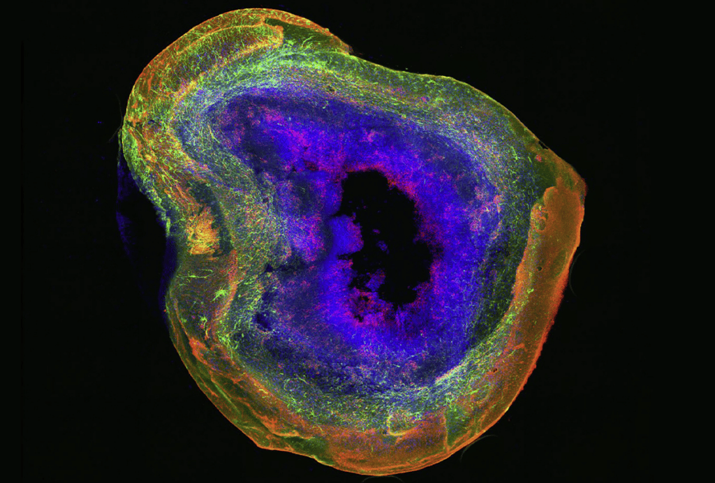
As neural stem cells mature into neurons, developmental genes move from the edge of the cell nucleus toward structures called nuclear speckles—a shift associated with increased gene activity, a study of developing human brain tissue found.
The findings suggest that a gene’s position inside the nucleus can influence whether it remains silent or becomes active.
Scientists have traditionally focused on chemical tags and regulatory proteins as the main controls on gene activity, says Yin Shen, professor of neurology at the University of California, San Francisco, who wasn’t involved in the study. Finding that where a gene sits inside the nucleus may also matter offers a new way to study normal brain development and neurodevelopmental conditions, she adds. “This work shifts the way we think about epigenetic regulation.”
Mutations in chromatin-regulating proteins are commonly associated with autism and other neurodevelopmental conditions. Yet conventional traits such as histone modifications and DNA accessibility often fail to explain the resulting changes in gene activity, says study investigator Daniel Lim, professor of neurological surgery at the University of California, San Francisco.
“We felt that we were missing something,” Lim says. So, the team began investigating whether a gene’s position inside the nucleus could provide that missing layer of information.
A
bout 30 to 40 percent of the genome is anchored to the nuclear lamina, a protein mesh lining the nucleus’s inner edge, where genes tend to be less active. By contrast, genes associated with nuclear speckles—small structures inside the nucleus that concentrate proteins involved in transcribing and processing RNA—tend to be highly active.Lim and his colleagues mapped which parts of the genome were associated with the nuclear lamina and speckles in specific cell types from human cortical tissue at 17 and 20 weeks of gestation. As radial glia—the stem cells that give rise to many neurons in the cerebral cortex—matured into neurons, about 23 percent of the genome moved either toward or away from the nuclear lamina.
Of the 739 genes that detached from the lamina, about 41 percent moved to nuclear speckles. Among these genes are several involved in brain development and neuronal communication, such as SATB2, MEF2C and SYT1, which have been linked to neurodevelopmental conditions.
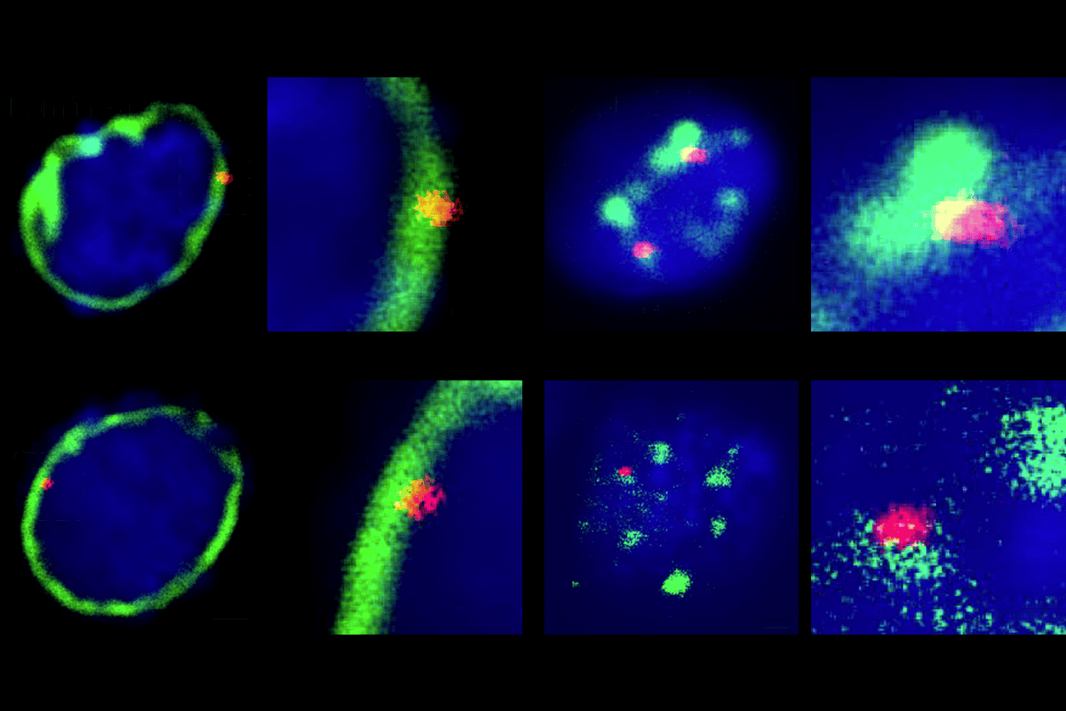
Microscopy confirmed the movement of five neuronal genes. In a brain region rich in radial glia, the genes tended to sit near the nuclear edge, whereas they appeared farther from the lamina and closer to speckles in brain regions where mature neurons accumulate.
The team then focused on developmental genes carrying both an activating and a repressive chemical mark, a combination that is thought to keep them silent but ready to switch on. During neurogenesis, genes that lost the repressive mark became much more actively expressed if they also moved away from the nuclear edge, whereas genes that stayed at the lamina changed little. And when the researchers experimentally removed the repressive mark, genes that remained at the lamina still stayed quiet. This suggests that location helps determine whether these poised genes are fully switched on.
The results indicate that the nuclear edge suppresses gene expression partly by keeping the genes away from the machinery needed to complete transcription. The team reported the findings last month in Nature.
S
hen compares earlier knowledge of nuclear organization to knowing that a city has different neighborhoods but lacking “a detailed address book showing where each resident lives.” The work shows that understanding where genes reside within the nucleus is crucial, she says.The study is an important step toward showing that a gene’s three-dimensional position in the nucleus can influence its activity, says Aleksandra Pękowska, who leads the Dioscuri Center of Chromatin Biology and Epigenomics at the Nencki Institute of Experimental Biology in Warsaw, and who wasn’t involved in the work. But, she says, “the key experiment will be to establish causality,” including testing whether restoring a gene to the correct nuclear position can rescue its expression independently of local chromatin marks.
Future work should also explore factors that could influence gene movement, including mutations in chromatin regulators, Pękowska says. She adds that the implications may extend to laminopathies, in which disrupted nuclear architecture is associated with transcriptional abnormalities.
The findings may also have implications for neurodevelopmental conditions, says Hongjun Song, professor of neurological sciences at the University of Pennsylvania, who wasn’t involved in the study. Because several genes that move between the nuclear lamina and speckles are involved in brain development, disrupting that movement could contribute to such conditions, Song says.
The work, he adds, “offers a new perspective on disease mechanisms and [may] eventually help identify therapeutic targets.”