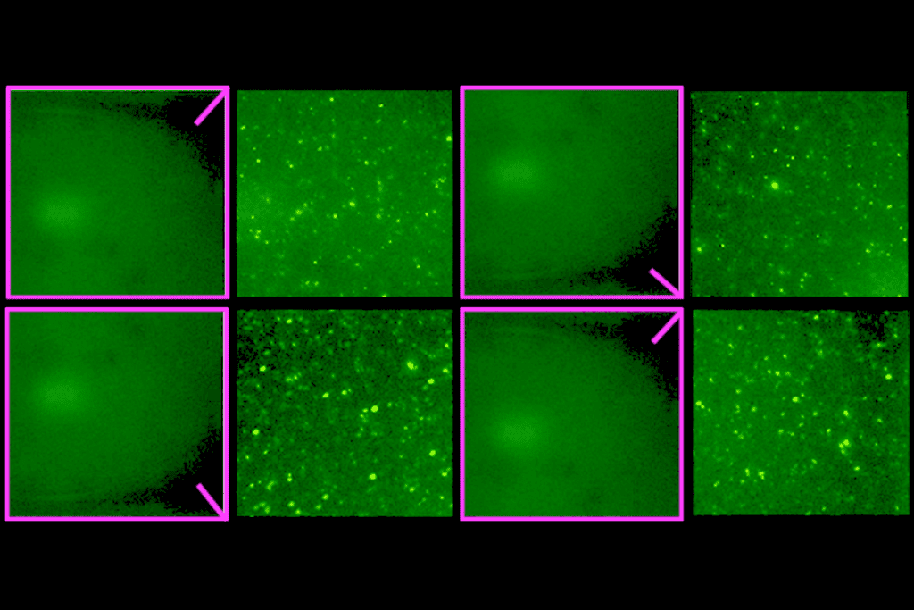
Circuit transformer: A three-day-old larval zebrafish’s visual experiences remodel the interneurons that determine the retina’s output, ultimately transforming how the animal engages with its surroundings.
Courtesy of Paride Antinucci
Add us as a Preferred Source on Google

Early experience reshapes zebrafish retinal cells, alters behavior

The cells undergo a classic form of plasticity previously thought to occur only in downstream visual circuits.

By Siddhant Pusdekar
13 August 2026 | 0 min watch

Visual experiences during early life famously shape cortical circuits. Animals deprived of vision in one eye rewire their cortex to favor the other eye, according to Nobel Prize-winning work by Hubel and Wiesel. And animals raised in controlled visual environments—surrounded by vertical stripes or horizontal ones, for example—adjust their cortical neurons’ orientation tuning.

This kind of experience-dependent plasticity also occurs in the retina itself, leading to lasting changes in behavior, according to a recent zebrafish study in Neuron. The sensory structure had long been thought to be hardwired.

“The field in general doesn’t think that activity has any effect on the retina, and it’s always [acting] downstream,” says Marla Feller, professor of neuroscience at University of California Berkeley, who wasn’t involved in the study.

Previous research suggests that waves of spontaneous neuronal activity in the retina that begin prenatally and continue till mice open their eyes help shape visual circuitry. The new work is the first to show that what an animal sees can prompt retinal activity that changes the shape and function of its interneurons, altering downstream processes including behavior, Feller says.

Vertebrates share many aspects of wiring in the retina, where layers of interneurons transform the pixel-like input from photoreceptors into distinct information channels encoding features of the visual environment. This commonality includes amacrine cells, which are one of the most diverse kinds of interneurons, says Robert Hindges, professor of developmental neurobiology at King’s College London and an investigator on the new study.

In zebrafish, amacrine cells expressing the cell adhesion molecule teneurin-3 respond to visual stimuli oriented parallel to the cells’ physical orientation, Hindges and his colleagues reported in 2013, and these cells are essential for establishing orientation selectivity. Mice and rabbits have similar amacrine cells that shape the orientation selectivity of retinal ganglion cells.

Amacrine cells, because of their role and the fact that they can be genetically labelled, seemed like a good place to start looking into the effects of visual environment on developmental plasticity in the retina, Hindges says.
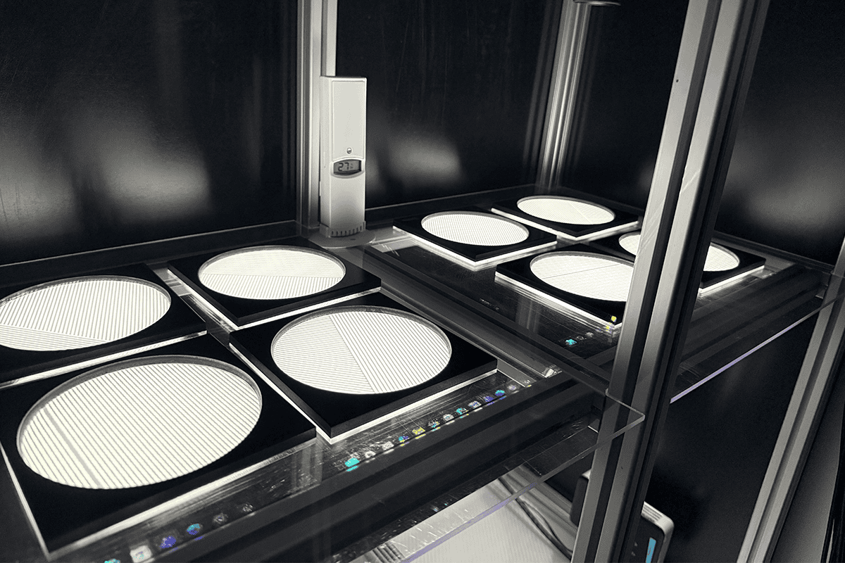

Zebrafish experimental setup.
Swim preference: Zebrafish preferred to swim into parallel-oriented stripes, but if they saw only horizontal stripes for the first five days after fertilization, they did not show a preference.

I

n the new study, zebrafish larvae lived in V-shaped channels with either horizontal or vertical black and white stripes on the walls for the first five days after fertilization.

Amacrine cells oriented parallel to the stripes became more elongated than usual, while others became rounder. Since these cells are distributed evenly across the retina, shape changes suggest that some occupy more space than others, says study investigator Phoebe Reynolds, postdoctoral fellow at the Friedrich Miescher Institute for Biomedical Research.

At this stage, orientation-selective retinal output to the optic tectum was biased towards the stripe orientation the fish saw, the researchers found, and the bias persisted for at least two days after the animals were moved to a neutral environment.

In another series of experiments, freely swimming fish given a choice between stripes of different angles preferred swimming towards stripes that ran parallel to their bodies, but those raised in a horizontal environment showed no preference. Fish lacking the TENEURIN-3 gene and raised in a horizontal environment, however, behaved normally, which suggests the animals can see the stimuli, but the loss of the gene “disturbs the cells in a way that they lose the ability to show plasticity,” Hindges says.

It is unclear why only the zebrafish raised in a vertical stripe environment prefer parallel-oriented stripes, he says. In addition to exploring the behavior further, Hindges says he is interested in looking at how “the changed amacrine cells” connect differently to ganglion cells.

The study is novel because it shows that “you can raise animals in a very specific environment, and their retinal circuits have now changed so that they’re better for that environment,” says Alexandre Tiriac, assistant professor of biological sciences at Vanderbilt University. But this may be more important in fish than in other animals because they “begin exploring their environment very early in their development,” he adds.

In mice, the effects of spontaneous retinal waves are known, but the existing evidence had not explained how activity shapes development and function, Tiriac says. By linking morphology, function and behavior, this study fills a much-needed gap in the field, he says.

Feller says the shape changes to the amacrine cells are particularly intriguing. It means it is now possible to study “what in the cell reads out that activity pattern and changes either synaptic strength or morphology,” she says. “We all want to understand what all of these things mean in terms of interpreting the natural world.”

Sign up for our weekly newsletter.

Catch up on what you missed from our recent coverage, and get breaking news alerts.