Brain mapping efforts, so far dominated by transcriptomics and connectomics, are putting a spotlight on lipids of late.
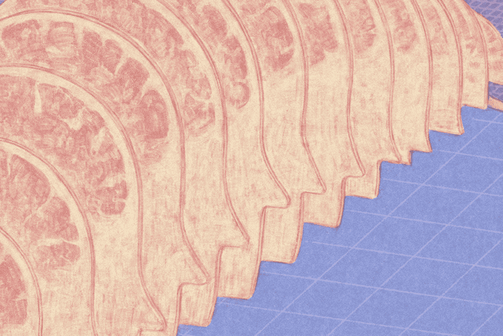
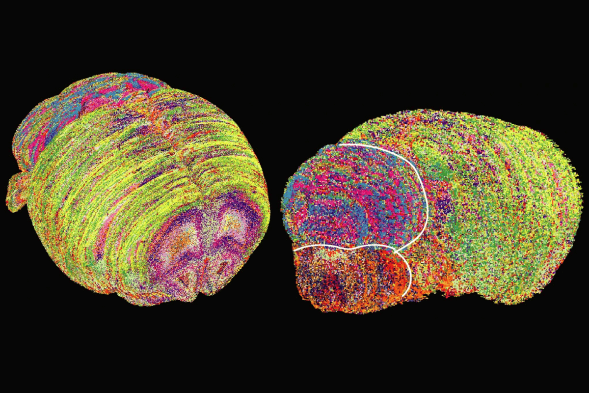
A new atlas described last month in Nature lays out the spatial distribution of 172 membrane lipids across 109 brain sections of 11 mice, with unprecedented resolution. Lipids cluster into 539 distinct regions, dubbed “lipizones,” and paint a picture of brain structure that is as intricate as, and distinct from, what is visible on gene expression maps.
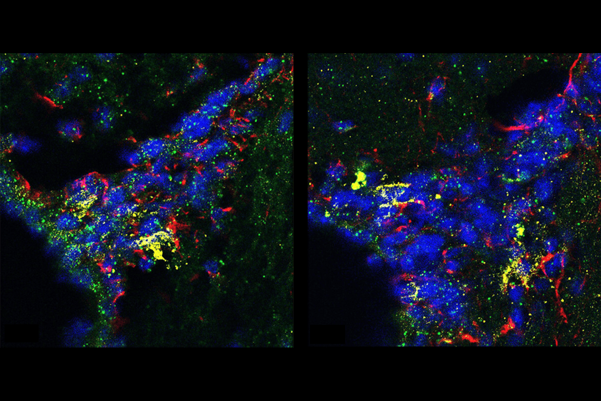
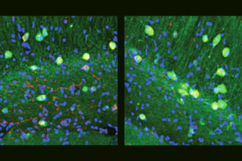
A temporal trace of brain lipids, published in July in Science Advances, shows that they change in quiescent neural stem cells, which are in a reversible state of nonproliferation. As mice age, these cells shift toward lipids with polyunsaturated fatty acid (PUFA) side chains that are highly unsaturated, and, as a result, the cells have less ordered membranes. “This could be related to their functional decline,” says study investigator Xiaoai Zhao, assistant professor of comparative medicine at Yale School of Medicine.
The two studies represent important progress in an otherwise understudied field of neuroscience, says Bruce Cohen, Robertson-Steele Professor of Psychiatry at Harvard Medical School, who was not involved in either study.
“Genes and proteins are the prom kings and queens” of neuroscience, he says, but lipids deserve attention, too: They constitute the membranes of cells, organelles and the nucleus; they provide energy, serve as signaling molecules and, in people with late-onset Alzheimer’s disease, become altered in astrocytes.
“Anything that goes on with a cell goes across a membrane, along a membrane, within a membrane,” Cohen adds. “Most of us aren’t adequately exposed to the importance of lipids, and we should be.”
O
The new brain atlas, created using mass spectrometry imaging, does not map neatly onto other ones, which trace the RNA expression of lipid-producing enzymes, metabolism-related genes or the full Allen Brain Atlas transcriptome, the team found.
For La Manno, that was a surprise, he says, and he recalls stating in an “overassertive tone” early in his Ph.D. program, “If there is something that changes in a cell, you should be able to see it in the RNA because [the gene] is where everything comes from.”
“Flash forward some years,” he says, and he found himself looking at, in a cortical region involved in fine-grained whisker sensation, 14 distinct lipizones not distinguishable by transcriptomics alone. “You cannot, simply by looking at gene expression, understand the story.”
The new lipid map also reveals a “shadow of connectivity,” he says, because of the myelin sheaths of long-range neurons and the lipid footprint they leave. “We start to see basically the same lipidome, millimeters away, in different regions. The lipids let us see connectivity in ways that we were not [able to see before].”
The difference in lipids between male and female mice was less dramatic than that between mid-gestation pregnant females and nonpregnant females. As female brains remodel during pregnancy, most lipizones are affected, with some changing size but not composition, and others changing composition but not size. The cortical regions most affected suggest that those lipid changes tune sensory circuits, perhaps to increase sensitivity to their pups. The enzymes that synthesize these lipids are controlled by master regulators that in turn receive signals from glucocorticoid receptors that sense stress hormones, showing how hormones could orchestrate this lipid tuning.
Specific enzymes track with age-related lipid changes, too, Zhao and her team found. Five enzymes fit two of these three criteria: They remodel PUFA side chains, they are highly expressed in quiescent neural stem cells, or their expression significantly changes with age.