Microglia are professional phagocytes, surveying the brain and cleaning up neuronal debris, damaged proteins and other biomolecules. The cells can also make contact with axons to scoop out large organelles containing degraded proteins, a new preprint shows.
The study reveals a novel exit route for debris that neurons could call upon when needed, says Ralph Nixon, professor of psychiatry and cell biology at New York University, who was not involved in the work.
Neurons can eject their garbage into extracellular spaces. They can also package waste generated at presynaptic active zones into organelles and traffic them to the soma for degradation. Occasionally, in stressed or active neurons, these proteolytic organelles can get too bloated to make it through the axon, says study investigator Shigeki Watanabe, professor of cell biology at Johns Hopkins University. The study, posted on bioRxiv in July, suggests microglia might be recruited to get rid of this buildup.
After observing microglia take up organelle aggregations from neurons in a cell culture, the researchers began looking for the same thing in a live brain, Watanabe says. Outside healthy brains, microglia have an “amoeba-like” appearance and behave differently, he says. “We cannot publish anything without in-vivo imaging in microglia.”
This work involved years of trial and error, because “you’re kind of imaging blindly,” says study investigator Renee Pepper, a postdoctoral researcher in Watanabe’s lab.
I
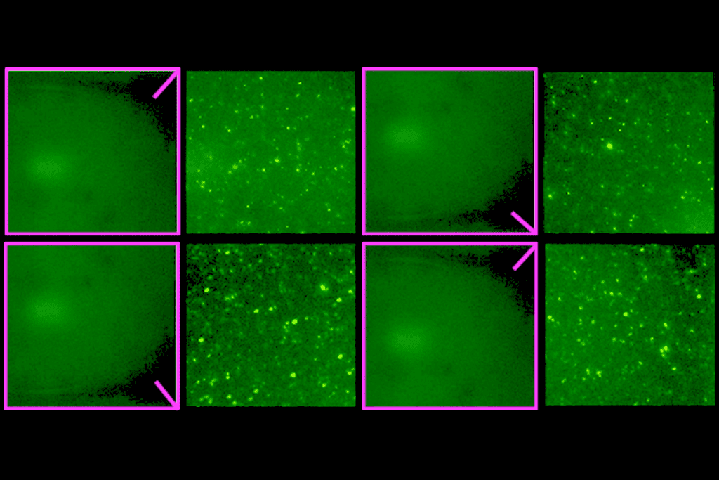
n the somatosensory cortex, microglia extended processes toward an average of 77 percent of the observed stationary organelles during 15-minute two-photon imaging sessions. Over several minutes, processes would “touch the organelle and pull back, and then go back again,” Pepper says, and following those contacts, a fluorescent tag on the neurons’ proteolytic organelles moved to the microglia.These interactions, which the researchers call skoupocytosis, are most common in the axon and appear to be distinct from previously documented instances of microglia nibbling synapses.
The protein makeup of synaptic terminals from artificially stimulated neuronal cultures revealed high levels of an enzyme called alpha/beta hydrolase domain-containing (ABHD16A). It regulates the amount of an extracellular form of phosphatidylserine (PS), a signaling molecule involved in microglial synaptic pruning. Blocking the enzyme reduces the proportion of PS outside the cell and increases the size of organelles accumulating in neurons, the new study shows.
This suggests that PS, once released outside the cell, could act as a flare for microglia to perform skoupocytosis, and that microglia don’t indiscriminately engulf bits of neurons but know precisely what to take, says Nicole Scott-Hewitt, assistant professor in the cell biology department at Duke University, who was not involved in the study. The new study, Scott-Hewitt says, adds to our understanding of the language of molecules that neurons and microglia use to communicate with each other.
Although the findings hint at a mechanism, the details have yet to be worked out, and there are likely more molecules involved, Watanabe says. How microglia interpret neuronal distress signs correctly is what he would like to look into next, he says.
A strength of the study is combining in-vivo, in-vitro and in-situ approaches, along with imaging techniques, says Marie-Ève Tremblay, professor of medical sciences at the University of Victoria, who was not involved in the study.
Observing microglia in the healthy brain is especially tricky, Tremblay says, because they “rapidly transform on a timescale of seconds and minutes, and can become toxic when taken out of their brain environment.” The new study expands on the past two decades of in-vivo work in showing that microglia are “extremely dynamic in the healthy brain,” she says.
Skoupocytosis might be rare in healthy brains, however, because the proteolytic organelles produced in healthy states are typically small enough to squeeze through the axon, Nixon says. Although the researchers found skoupocytosis in vivo without manipulating neuronal activity, the only way to reliably get the organelles to accumulate for in-vitro experiments was by artificially inducing activity, Pepper says. She plans to look at how synaptic activity recruits the molecular players involved in skoupocytosis and how important it is for synapse function.
“Seeing two things that are different, like interacting in a coordinated” way stimulates an “innate biological curiosity,” says Fernando González Ibáñez, who was not involved in the study and who asked to be identified as a Mexican postdoctoral researcher in Tremblay’s lab. “Taking something out without this cell bursting,” is a tricky process involving a host of mechanical forces, he adds.