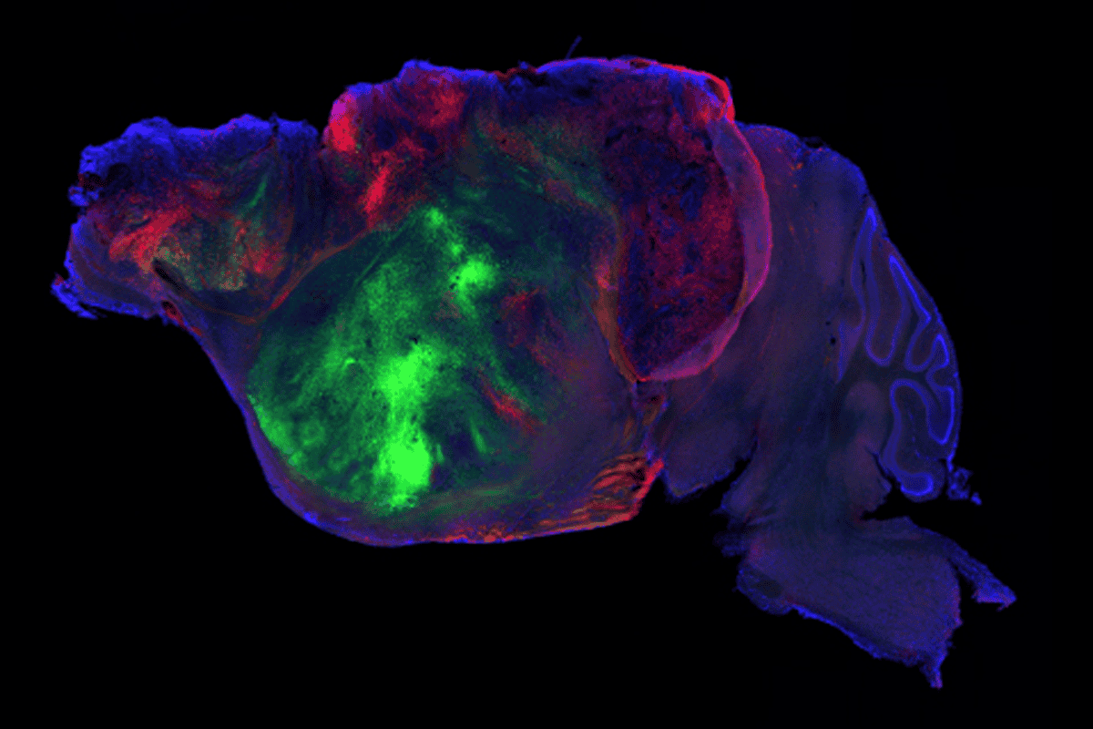
Nerve fibers from the human graft in mouse brain.
Long range: Nerve fibers from the human graft (green and red) extend throughout the mouse brain (blue).
S. Pașca lab, Stanford University
Add us as a Preferred Source on Google

Human brain organoids flourish in emptied mouse cortex

The chimeric mice could help untangle the biology of human brain development or conditions such as autism.

By Brianna Abbott
16 September 2026 | 6 min read

Human cortical organoids transplanted into newborn mice integrate into the nervous system and form circuits that support electrical activity, according to a new study published today in Nature. The mice were engineered to lack nearly all of their cerebral cortex, creating space for the human-derived tissue to take hold.  

The researchers then used the ‘xenocortical’ mice to model hypoxia, a state that occurs when brain cells don’t get enough oxygen. When hypoxia occurs prenatally or around the time of birth, it can cause cerebral palsy; it is also associated with autism and epilepsy.

The transplantation approach could serve as a new tool to study typical development and neurodevelopmental conditions, as well as test potential therapeutics, says Hongjun Song, professor of neuroscience at the University of Pennsylvania’s Perlman School of Medicine, who was not involved in the study. 

“Now you have a model, a live model where you can actually test all of this with human cells in live animals,” Song says. 

The lack of access to living brain tissue has presented a long-standing barrier to understanding human development. Brain organoids derived from human stem cells have opened up fresh possibilities, and transplanting the organoids into animals such as rats can help the human cells mature even further and form more complex circuits. But physical space within the rodents is finite, and the human neurons can struggle to establish themselves. 

“They will be outcompeted by mouse cells very quickly,” says Sergiu Pașca, professor of psychiatry and behavioral sciences at Stanford University and the study’s senior investigator. “It’s also very difficult to actually distentangle, to be honest, the effect of the human versus the mouse.” 

In the new paper, Pașca and his team used a knockout strategy to design a mouse model. They deleted ESCO2, a protein that aids in cell division, from cells expressing a marker for the dorsal and medial pallium, the areas of the embryonic forebrain that give rise to the cortex and hippocampus. The team also designed the mice to be immunocompromised so that the animals would not reject the human cells. 

The resulting mice were missing 98 percent of their cortex and hippocampus and had a 50 percent decrease in total brain tissue, compared with controls. Even so, the mice exhibited only minor behavioral changes, such as having a more cautious gait. 

“These animals are not perfectly normal but also surprisingly functional, much more than we thought they would be,” Pașca says. 
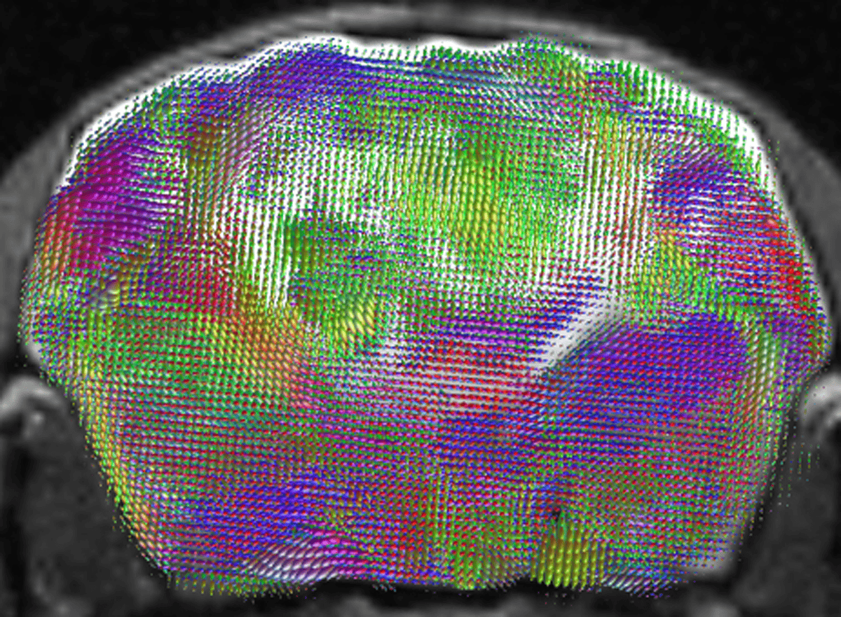
MRI scan of an estimated map of nerve-fiber pathways.
Road map: An MRI scan reveals an estimated map of nerve-fiber pathways and directions (green, top to bottom; red, front to back; blue, side to side).
S. Pașca lab, Stanford University

T

he researchers successfully transplanted the human organoids into 25 out of 29 newborn engineered mice. The human-derived tissue proliferated, taking over the space and growing 4.7-fold between two and three months after transplantation. The grafts integrated into the mouse nervous system, and the mice performed similarly to wildtype controls on motor, learning and social tests. 

“Replacing a substantial portion of an animal brain with human neural tissue represents an impressive technical advance,” In-Hyun Park, associate professor of genetics and neuroscience at Yale School of Medicine, wrote in an email to The Transmitter. Park was not involved in the study.

The grafts contained a diverse range of human cortical cell types, including rare von Economo neurons. VENs, which appear in large-brained, social animals, are implicated in some neuropsychiatric conditions and have been challenging to produce in cell culture, the paper says. 

The researchers then tracked the calcium dynamics in graft-derived neurons. Large, synchronous bursts occurred every few minutes, correlating with mouth and facial movements. Electrophysiologic signals also matched the bursts and were detected across the electrode, suggesting that the graft forms into a functional neural network, the researchers wrote.

To test if the mice could be used to study disease, the researchers modeled hypoxic injury, which affects humans more severely than mice. The team exposed xenocortical, cortex-less and control mice to low oxygen for five hours. The xenocortical mice were uniquely impacted, showing trouble maintaining their balance and sustaining a steady gait. 

T

he grafted organoids have their own set of limitations, Pașca says. The human-derived neurons still develop at their same, slower pace than the mouse cells. The tool so far applies only to the earliest stages of human development and is limited by the lifespan of the mice.

The neurons also aren’t organized in layers, and the organoids lack inhibitory neurons. Variability in how the organoids grow and integrate might make it difficult to answer some questions, says Joseph Gleeson, professor of neuroscience at the University of California, San Diego, who was not involved with the work.

“It doesn’t build a new cortex. What it does is it gives a lot of room for these organoids, or the cells in the organoids, to divide and grow and take up residence,” he says. “It’s the single biggest finding and also the single biggest limitation.”

“This was about the best you can do at this point,” he adds. “I’m really interested to see where the field goes.”   

The team also published details on the experiment’s ethical oversight, describing how they consulted with bioethicists and other neuroscientists throughout the project. In addition, Stanford convened a group of legal scholars, patient advocates, ethicists and scientists to oversee and provide feedback on the experiments, as neuroscientists continue to grapple with the ethics of creating increasingly sophisticated human brain organoids. 

Meanwhile, the model could help investigate both environmental injuries and genetic conditions. “Every time you have somebody with a genetic mutation, you can either induce it in control cells, or you can take that patient’s cells and transplant them,” Pașca says. 

The approach is also apt to answer basic questions about how human circuits form, he adds. “What makes human circuits unique? Or primate circuits so unique?” he says. “How are they processing information very early in development?” 

Sign up for the weekly Spectrum newsletter.

Stay current with the latest advancements in autism research.