All the dorsolateral prefrontal cortex is a stage and all the genes merely players; they have their exits and entrances across three acts divided by age, according to a new human brain-cell atlas published today in Nature along with eight companion studies.
Turbulent changes related to neurogenesis and gliogenesis make for a dramatic first act leading up to adulthood. The second act, from early adulthood through middle age, is eerily silent, but a new cast of characters dominated by glia takes the stage for the third act, at around age 60, according to the atlas, which shows how gene activity changes in individual cells of the human prefrontal cortex over the span of a lifetime.
The data are publicly available and come from 1,494 people, who ranged in ancestry, health and psychiatric profiles and age, from before birth to more than 100 years. The studies reveal the nonlinear dynamics of disease progression and weakened clock-gene rhythms in people aged 60 and up, among other novel findings.
The work is a “herculean effort” that is “transformative for our understanding of the prefrontal cortex across the human lifespan, in neurodegenerative and psychiatric disease, and its genetic regulation,” says Jennifer Below, professor of genetics at Vanderbilt University Medical Center. Below was not involved in the work but reviewed one of the companion papers.
The atlas is “the largest resource with cellular resolution released to date,” Below adds. Comparable datasets containing single-cell or single-nucleus transcriptomics have focused on specific age groups or brain disorders, or have smaller cohorts.
“Studying disorders separately can obscure shared biology, while combining datasets generated using different methods can introduce technical differences that resemble disease effects,” says study investigator Panos Roussos, professor of psychiatry and of genetics and genomic sciences at the Icahn School of Medicine at Mount Sinai.
The new dataset includes 500 people diagnosed with Alzheimer’s disease, 112 with diffuse Lewy body disease, 85 with vascular dementia, 40 with Parkinson’s disease, 47 with tauopathy, 15 with frontotemporal dementia, 120 with schizophrenia and 55 with bipolar disorder. Many of these people had multiple diagnoses.
R
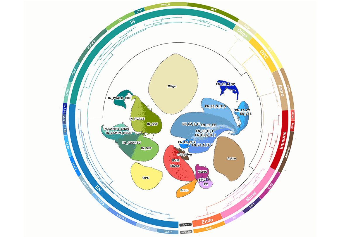
oussos and his colleagues in the PsychAD Consortium isolated single nuclei from dorsolateral prefrontal cortex tissue and conducted multiplexed single-nucleus RNA sequencing. They focused on the dorsolateral prefrontal cortex because it “contributes to working memory, planning, decision-making and cognitive control—functions affected across many of the disorders we studied,” he says.Based on genetic markers, they sorted the 6.3 million nuclei by cell type, including excitatory and inhibitory cells, astrocytes, immune cells, oligodendrocytes, oligodendrocyte progenitor cells and vascular support cells.
Cell type accounts for 50.5 percent of the variation in the dataset; the second-largest source of variation was person-to-person differences. This result shows that “nuclei from the same person are related observations,” so researchers planning to use the dataset should avoid treating “each nucleus as an independent person,” Roussos says.
Non-neuronal cells are more abundant than usual in the brains of people with neurodegenerative disorders, whereas deep-layer excitatory neurons are more abundant in people with neuropsychiatric disorders, according to an analysis using a custom statistical tool. The groups also differentially expressed some of the same genes, a different statistical tool demonstrated. Many of those genes are essential for cell function.
Different cell types also follow specific trajectories over the course of disease progression, a neural network model revealed. This is a novel perspective, showing that gene-expression patterns change nonlinearly across disease progression and can even impact it, says Michael Lutz, professor of neurology and pathology at Duke University, who was not involved in the work but reviewed the paper.
The broad age distribution enabled the researchers to look at transcriptomic changes through development and aging. They found “extensive developmental changes, followed by relative molecular stability through much of adulthood and renewed changes in later life, particularly in glial support and immune cells,” Roussos says.
Clock genes associated with circadian rhythms are particularly dysregulated in people aged 60 or older, an analysis of data from approximately 200 neurotypical adults revealed. These patterns were reconstructed from donors with different times of death, so they help “generate hypotheses about circadian aging rather than directly measuring changes in an individual’s sleep,” Roussos says.
T
he dataset also represents people with different ancestries and underlying genetic traits, which the researchers mapped onto observed gene-expression patterns and built models of how genetic variation shapes gene regulation. This pointed them to “new targets and potential mechanisms of risk that can be exploited to develop better drug therapies,” Below says.However, “many groups were not represented, and there remains a lot of opportunity to expand such studies across populations and exposures,” Below says.
It is the first time this kind of analysis has been applied to people of non-European descent at this scale, says Seth Ament, professor in the Department of Psychiatry and Institute for Genome Sciences at the University of Maryland School of Medicine, who was not involved in the study. The analysis included 439 people of non-European or mixed ancestry. It shows convergence across ancestry for “cell-type-specific mechanisms, some of which we didn’t know about,” he says.
The brains were collected postmortem, “and what’s present in that brain reflects the entirety of someone’s life experience in some ways,” says Ament, who reviewed the paper. For complex diseases, such databases are mainly useful for generating hypotheses and pointing to where researchers should be looking, he says. Ament and his colleagues posted a preprint in July describing changes in single-cell gene expression across different ages, representing more than 2 million cells.
Next, Roussos says he wants to “expand across brain regions and populations, integrate gene expression with other molecular measurements, and test the mechanisms suggested by the atlas.”
Lutz says he plans to use the database in combination with other multi-omics atlases, such as Rush Memory and Aging Project and the Alzheimer’s Disease Neuroimaging Initiative dataset, for orthogonal validation and to gain deeper insight into disease mechanisms.